
Fetal urological aspect: Fetal Genitourinary Disease 2015
Educational content from recorded physician discussions — not medical advice. Talk to your (or your child's) care team about your situation.
Video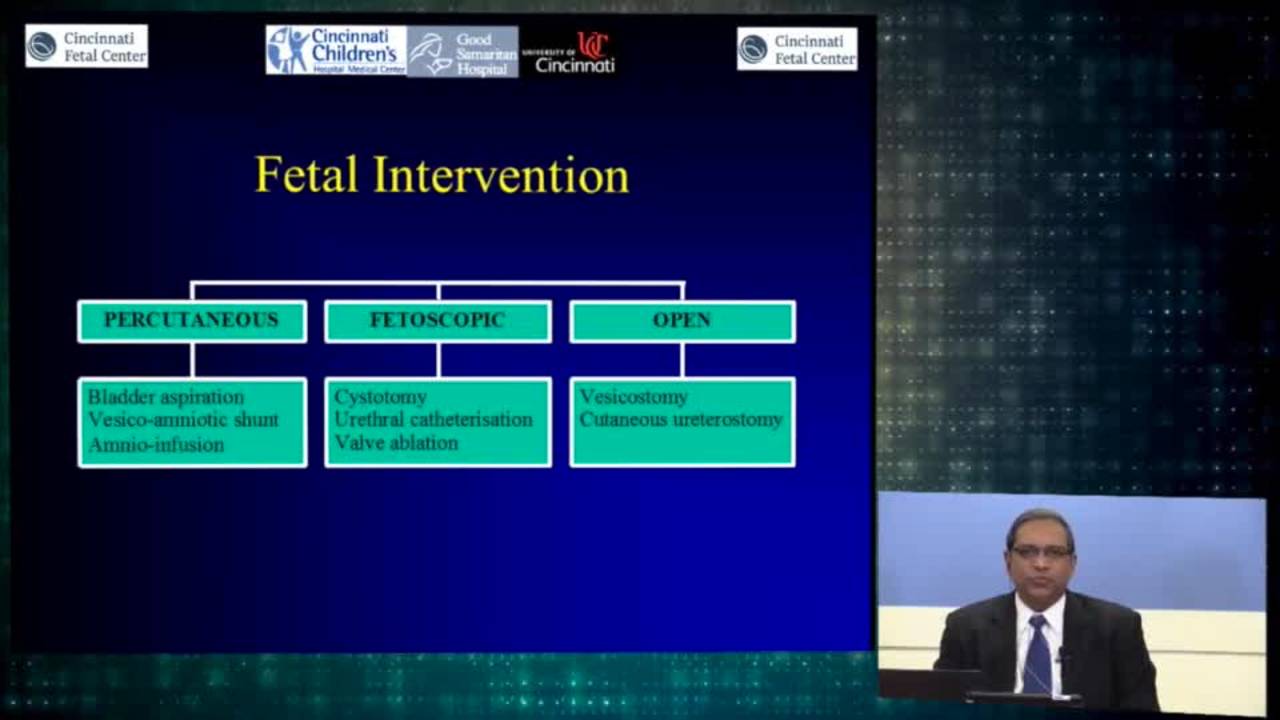
Urologic Fetal Intervention: Cincinnati Fetal Center
61 min · Published Nov 2018
Video
Prenatal diagnosis of fetal lower urinary tract obstruction: Fetal...
40 min · Published Jul 2017
Video
Overview of Prenatal Diagnosis: Cincinnati Fetal Center
40 min · Published Nov 2018
Video
Renal transplantation: Fetal Genitourinary Disease 2015
13 min · Published Jul 2017
Video
Urologic intervention: Cincinnati Fetal Center
28 min · Published Nov 2018
Video
Fetal Diagnostic Imaging: Cincinnati Fetal Center
10 min · Published Nov 2018
Video
Pediatric Surgical Oncology Research Collaborative (PSORC): Studying Rare Pediatric Tumors
56 s · Published May 2026
Video
Update Course Rewind 2025: Hirschsprung + ARM: Rare but Real
1 min · Published May 2026
Video
Update Course Rewind 2025: Hirschsprung + ARM: Rare but Real
1 min · Published May 2026
Video
Pooling Patients to Study Rare Pediatric Tumors: An Introduction to PSORC
56 s · Published May 2026
Video
The fetal frontier: A review of current and emerging fetal therapies for genetic diseases
44 s · Published May 2026
Video
Indocyanine green assists with sentinel lymph node mapping in pediatric and adolescent patients
1 min · Published May 2026
What the experts said
Not all fetuses with urinary tract obstruction are candidates for intervention; some are too healthy (normal AFI, non-obstructive dilation, unilateral UPJ obstruction) where intervention risk outweighs benefit.
Some fetuses are too sick for intervention to provide benefit (renal cystic dysplasia, abnormal urinary parameters, abnormal karyotype, multiple congenital anomalies).
Vesicoamniotic shunts have not lived up to their promise because they are long thin tubes with high resistance; while they allow urine reflux for lung development, they do not adequately relieve pressure causing upper tract injury.
In adults, bladder pressure exceeding 40 cm H2O (29 mmHg) injures kidneys and eliminates net glomerular filtration.
Safe bladder pressure thresholds for fetal kidney development are unknown and should be studied by measuring opening pressure during vesicocentesis.
Intravillous pressure that occludes venous flow in the placenta is about 23 mmHg; resting amniotic fluid pressure is 5-8 mmHg.
Shunts have provided pulmonary survivors but have not significantly improved renal outcomes; fetoscopic and open vesicostomy approaches that reduce bladder pressure to near-zero may offer better renal preservation.
When a shunt is placed and the bladder collapses, upper tracts (ureters, renal pelvis) significantly decompress to near-normal appearance.
Even after bladder decompression with shunts, residual hydroureteronephrosis persists in some cases, suggesting incomplete pressure relief.
Fetoscopic cystoscopy may be superior to shunting because it allows cyclical voiding (more physiologic drainage), determines etiology (PUV vs. atresia), avoids amnioinfusion complications, and avoids shunt migration/blockage.
Shunt complications include migration (can end up around the cord) and gastroschisis when placed at 17 weeks.
Bladder cycling enabled by fetoscopic procedures is important for bladder health, not upper tract protection; the main renal benefit comes from pressure reduction via a normal urethral lumen.
A 10% risk of urological fistula from laser ablation is preferable to renal dysplasia because fistulas can be surgically repaired postnatally with minimal morbidity.
Posterior urethral valves can sometimes be bluntly ablated by pushing a catheter through with a guide wire because the valves are flimsy.
Hydrodistention alone is unlikely to rupture posterior urethral valves; if it could, the pressure in the obstructed urinary tract should already blow them open.
Three serial bladder taps are not necessary if the first and second show favorable values (at or below threshold on first, clearly below on second); additional taps risk bladder rupture and ascites.
Fetoscopic procedures can be performed with epidural plus IV sedation and local anesthetic rather than deep general anesthesia, reducing maternal anesthetic risk compared to open fetal surgery.
Fetal anesthesia for fetoscopic procedures uses intramuscular vecuronium, atropine, and fentanyl to prevent fetal movement and mitigate pain response.
Fetoscopic cystoscopy technique uses a 3.3mm outer sheath fetoscope (1.2mm scope) with side port for guide wire, sometimes switching to a 4.9 French flexible ureteroscope for better maneuverability to access the bladder neck.
The PLUTO trial removed bladder taps from their protocol, but the trial had many flaws including lack of patient selection based on urine parameters.
The first bladder drainage reflects urine that has been sitting in the bladder and is not predictive; the second is also not predictive; the third provides fresher urine with better correlation to underlying renal injury.
Cincinnati Children's Hospital experience with open fetal vesicostomy: 6 cases offered to families with anhydramnios, 4 fetal deaths (all preterm deliveries), 2 survivors. One 5-year-old has normal creatinine and bladder function with solitary kidney; the other required transplant.
Open fetal intervention predisposes to very shortened gestational age (about 10 more weeks maximum) and the operated uterus is highly unstable with high risk of preterm labor.
There is no role for open fetal surgery for LUTO; offering the highest-risk procedure to the sickest patients exposes mothers to maximum harm for minimal fetal benefit and increases prematurity risk that further compromises already-damaged kidneys.
Open fetal vesicostomy should be considered only in patients with good prenatal prognostic profile who have failed fetoscopic intervention, not in the worst-prognosis patients.
In long-term follow-up (5-12 years) of shunted LUTO patients, 92% survived, 45% had normal renal function, 22% had mild insufficiency not requiring intervention, but 33% developed end-stage renal disease requiring transplantation.
Among long-term survivors, 67% could spontaneously void, while 33% required intermittent catheterization or continuous catheterization.
Time to transplant varies by diagnosis: posterior urethral valves at 10-12 years, prune belly/urethral hypoplasia at 4.5 years, urethral atresia earlier.
Children who progressed to end-stage renal disease had acceptable discharge creatinine but developed progressive injury from repeated infections, severe reflux, and valve bladder dysfunction over subsequent years.
Infants with LUTO triple their birth weight in the first year, revealing true renal reserve; discharge creatinine reflects a 3-5 kg body mass, but by age 1 they are 10-15 kg, generating 2-3 times more creatinine and potentially overwhelming marginal kidney function.
Creatinine at hospital discharge is misleading; creatinine at age 1 year is a better prognostic indicator. If creatinine at age 1 is less than 1.0, the child likely will not need renal replacement therapy; if above 1.0, there is high probability of needing dialysis or transplant.
The bladder initiates upper tract damage and is often underestimated; even with minimal kidney injury, concentrating defects cause polyuria, which damages the bladder, raises storage pressures, and creates a vicious cycle of progressive renal injury.
Patient selection is the most critical factor for any fetal intervention; early shunting experience included inappropriate candidates with aneuploidy and major anomalies.
Open fetal surgery for LUTO should only target fetuses with very high potential for success, not those with significant evidence of injury; the procedure has 4% loss rate and significant prematurity rate.
Fetoscopic cystoscopy needs proper evaluation to avoid repeating the mistakes of the PLUTO trial; just because a procedure can be done does not mean it should be done.
Cystoscopy altered the diagnosis in 25-33% of fetuses with suspected LUTO.
In a French/Brazilian/Houston series of 111 LUTO fetuses (60 no intervention, 16 shunted, 34 cystoscopy), both interventions improved 6-month survival, but only cystoscopy may prevent renal function impairment at 6 months.
In a Barcelona/Leuven series of fetal cystoscopy with laser valve ablation, urethra was accessed in nearly all cases, bladder size and amniotic fluid normalized in 80%, and among livebirths, none had pulmonary hypoplasia and three-quarters had normal renal function.
Fetoscopic cystoscopy creates a urological fistula in 9-10% of cases and causes recurrent severe LUTO in about 6%.
Open fetal vesicostomy was pioneered by Harrison in 1982 with fetal ureterostomies; he later reported 8 open vesicostomies with 100% technical success but 50% mortality, leading him to stop the procedure.