
Tips and Tricks: EA & TEF
With Dr. Steve Rothenberg · StayCurrentMD
Educational content from recorded physician discussions — not medical advice. Talk to your (or your child's) care team about your situation.
Video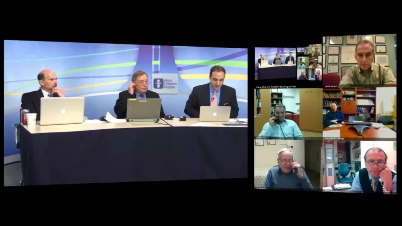
Leak after Esophageal Atresia Repair / TEF Repair: Discussion of World Experts
Dr. Todd Ponsky · 15 min · Published Dec 2012
Video
Thoracoscopic Repair of Tracheo-esophageal Fistula Tricks: Pediatric Surgery...
19 min · Published Jul 2017
Video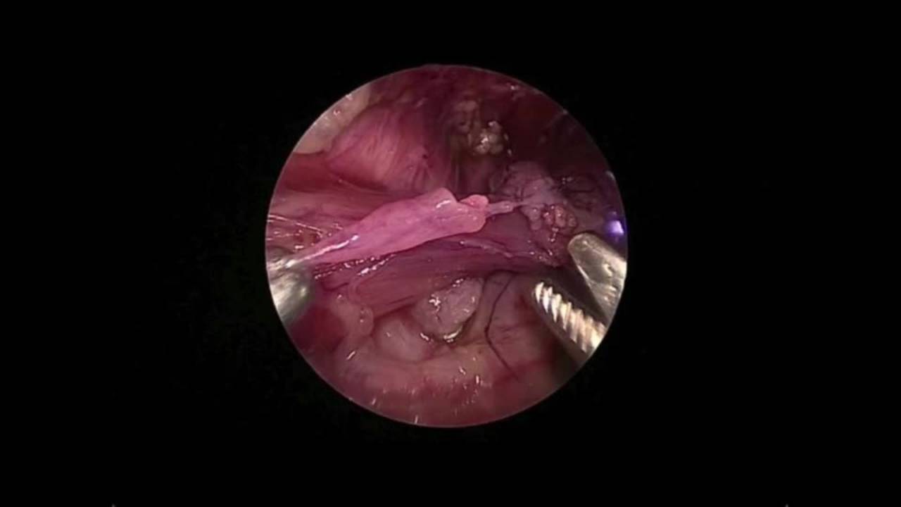
EA/TEF Discussion & Technique: Difficult Cases
19 min · Published Dec 2013
Podcast
Case Based Journal Review: Esophageal Atresia in 2022
17 min · Published Aug 2022
Video
Comparison of robotic versus thoracoscopic repair for congenital esophageal atresia
56 s · Published Feb 2024
Video
Glycopyrrolate for Anastomtic Dehiscence in Esophageal Atresia
2 min · Published May 2017
Only a few other public items share this expert — go deeper there →
Video
Pediatric Surgical Oncology Research Collaborative (PSORC): Studying Rare Pediatric Tumors
56 s · Published May 2026
Video
Update Course Rewind 2025: Hirschsprung + ARM: Rare but Real
1 min · Published May 2026
Video
Update Course Rewind 2025: Hirschsprung + ARM: Rare but Real
1 min · Published May 2026
Video
Pooling Patients to Study Rare Pediatric Tumors: An Introduction to PSORC
56 s · Published May 2026
Video
The fetal frontier: A review of current and emerging fetal therapies for genetic diseases
44 s · Published May 2026
Video
Indocyanine green assists with sentinel lymph node mapping in pediatric and adolescent patients
1 min · Published May 2026
What the experts said
Spitz uses a 6 French feeding tube (not a Replogle) across the anastomosis to feed the baby for the first few days after operation.
Jack leaves a 5 French feeding tube across the anastomosis, both for feeding and to protect the back wall during the anastomosis.
Steve uses blunt and sharp dissection when separating the upper pouch from the membranous trachea, only using cautery when he can clearly see the tissues and knows he is not injuring the membranous trachea.
Using cautery to dissect the plane between the membranous trachea and the medial wall of the upper esophageal pouch can result in holes in the membranous trachea, leading to an acquired tracheoesophageal fistula that is very difficult to fix.
Sharp dissection is the only safe way to dissect the upper pouch off the membranous trachea; cautery should never be used in that particular plane.
It is very hard to tie an extracorporeal knot with Vicryl suture.
If tying extracorporeally, PDS should be used instead of Vicryl because it slides better.
When there is an anastomosis under tension and concern about a leak, it is a good idea to get some tissue between the two suture lines to prevent development of a recurrent fistula.
A pleural flap or pericardial flap can be used to place vascularized tissue between suture lines.
In a baby, a thick pleural flap can be created that incorporates some of the muscle of the intercostal spaces and some fat.
The thing that causes most recurrent fistulas is a leak, and the thing that causes most leaks is too much tension on the anastomosis.
In premature infants with very small upper pouch and fistula, Steve's approach is to ligate the fistula thoracoscopically, mobilize the upper pouch if possible, and if unable to achieve anastomosis, place an internal stitch in the upper pouch under tension and return in 4-6 weeks for anastomosis.
The advantage of thoracoscopic approach is that without a big thoracotomy incision, the surgeon can go in, evaluate, and decide whether anastomosis is achievable.
Holger shares the experience that in premature infants, if only the fistula is ligated without dissection and the surgeon waits 4-8 weeks for spontaneous growth, the anastomosis can be performed later successfully.
If the azygos vein is ligated, the distal end can be opened and plastered onto the tracheal or esophageal suture line as tissue interposition.
Jack had a case where he placed only a clip on a fistula in a preemie without dividing it, and 3 weeks later the fistula recurred (recanalized).
If clipping a fistula, it must be divided; just putting a clip on can lead to recanalization.
When performing fistula division or ligation in a case where the two ends cannot be anastomosed, the distal segment should be tacked to the vertebral body (prevertebral fascia) with a stitch to prevent it from shrinking down.
More than 50% of long-gap EA cases are type C.
A paper by Upadhaya in the European Journal of Pediatric Surgery reported that ligating the azygos vein is associated with an increased rate of pneumonia.
David Vander says he never ligates the azygos vein.