
EA/TEF Discussion & Technique: Difficult Cases
With Dr. David Vanderzee & Dr. Jeff Blair · hosted by Dr. Todd Ponsky · StayCurrentMD
Educational content from recorded physician discussions — not medical advice. Talk to your (or your child's) care team about your situation.
Video
Thoracoscopic Repair of Tracheo-esophageal Fistula Tricks: Pediatric Surgery...
19 min · Published Jul 2017
Video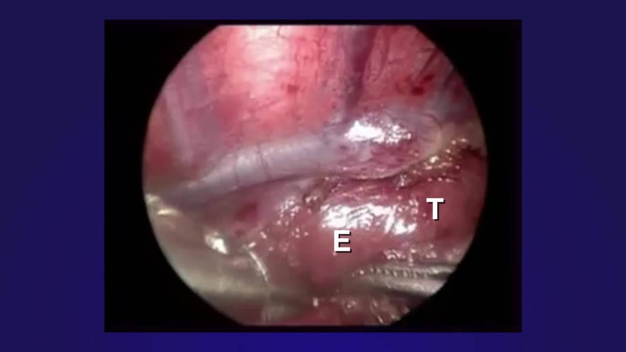
Thoracoscopic Repair of Esophageal Atresia with Distal Tracheo-esophageal...
5 min · Published Feb 2020
Video
Tips and Tricks: EA & TEF
Dr. Steve Rothenberg · 9 min · Published Dec 2012
Video
Thoracoscopic TEF Repair, Is It Really Better Than Open? Update Course 2018
18 min · Published Aug 2018
Video
Tricks - Imperforate Anus and Rectourethral Fistula
45 min · Published Nov 2018
Video
Update Course 2013: EA & TEF
38 min · Published Sep 2013
Only a few other public items share this expert — go deeper there →
Video
Pediatric Surgical Oncology Research Collaborative (PSORC): Studying Rare Pediatric Tumors
56 s · Published May 2026
Video
Update Course Rewind 2025: Hirschsprung + ARM: Rare but Real
1 min · Published May 2026
Video
Update Course Rewind 2025: Hirschsprung + ARM: Rare but Real
1 min · Published May 2026
Video
Pooling Patients to Study Rare Pediatric Tumors: An Introduction to PSORC
56 s · Published May 2026
Video
The fetal frontier: A review of current and emerging fetal therapies for genetic diseases
44 s · Published May 2026
Video
Indocyanine green assists with sentinel lymph node mapping in pediatric and adolescent patients
1 min · Published May 2026
What the experts said
For thoracoscopic EA/TEF repair, the gap between proximal and distal esophagus should be assessed preoperatively using X-ray with nasogastric tube and bronchoscope positioning to identify the fistula orifice.
Leaving one-quarter of the fistula uncut prevents cranial retraction of the distal esophagus and makes anastomosis easier.
Preserving one-fifth to one-quarter of the proximal esophageal tip provides a 'cap' for grasping without touching the anastomotic site.
If there is a 1-2 to 3 vertebral body gap, the assistant can pull the proximal esophagus caudally using the uncut cap.
The first stitch should be placed in the middle of the posterior wall rather than at the edge.
Optical trocars are used for initial port insertion in this technique.
The mucosa of the distal esophagus must be included in sutures to prevent postoperative stenosis.
6-0 PDS suture is used for the anastomosis.
The tracheoesophageal fistula is divided completely after 1-2 stitches are placed.
The uncut cap of the proximal esophagus is divided after 2-3 stitches are placed.
Todd Ponsky tried Yama's technique after seeing the video in September and found it worked well, though he was uncertain how much the traction contributed versus standard technique.
There is no downside to leaving the last bit of fistula attached—if the stitch sets up perfectly, it can be taken; if not, it can be cut.
David Vanderzee uses a transfixing suture to close the fistula rather than clips to ensure it doesn't come off.
David Vanderzee avoids clips because they tend to hook behind the suture being used for anastomosis.
For type C anastomosis with considerable length, David Vanderzee places two sutures and makes them into sliding knots to slowly bring the ends together, dividing tension between the two ends.
David Vanderzee finalizes the posterior anastomosis before placing a tube and closing the anterior wall.
Jeff Blair predicts that in the next decade, mechanical devices will be developed that can be passed through the upper pouch to mechanically grab and seal the lower pouch, possibly with thoracoscopic or imaging guidance.
Suet found the fistula much easier to identify and appreciate laparoscopically, allowing better assessment of how much esophagus to leave on the tracheal side.
Suet found dissecting the upper esophageal segment difficult, particularly in handling the esophagus without damaging the wall needed for anastomosis.
Todd Ponsky's experience with thoracoscopic TEF repair (3 cases over one year) has been that the first half during dissection and fistula division feels advantageous, but the anastomosis phase makes him question the approach.
Todd Ponsky has experienced a clip eroding into the esophagus.
Todd Ponsky uses metal clips, the same type Steve Rothenberg uses.
Weck Hema lock clips are used by many surgeons, but the applier is large and impairs visualization compared to metal clip appliers.
Weck Hema lock clips are difficult to remove if their position is unsatisfactory.
If the fistula clip is applied very tightly, it will crush the esophageal muscle and cause erosion, leading to recurrent fistula; the clip should just oppose the tissue rather than crush it.
Mark Woflan from Atlanta uses a technique where a stitch is placed through the chest wall, through the proximal end, through the distal end, and back out through the chest wall, holding the ends together during anastomosis.
Several instances have occurred where clips seem to erode or be implicated in recurrent fistula.
Steve Rothenberg uses clips routinely and has the largest experience, but has never had a clip erode.