13 views 0 likes

StayCurrentMD

GCMD Space · View profile →

Radiology: Pectus Innovations

Video Published 2019-01-11 Updated 2022-08-22

Timestops (5)

Topic Overview

A multidisciplinary discussion on preoperative imaging and cardiac assessment in pediatric pectus excavatum. The panel presents evidence that cardiac MRI has replaced CT as the preferred imaging modality, offering comprehensive anatomical measurement (Haller, correction, and depression indices), cardiac functional assessment, and screening for associated connective tissue abnormalities—all without radiation. Approximately 15% of patients demonstrate reduced right ventricular ejection fraction (<50%), particularly when the sternum compresses the RV free wall. The discussion addresses insurance requirements for objective metrics, the limitations of Haller index alone in patients with barrel-shaped chests, and emerging questions about optimal surgical timing and long-term cardiac outcomes.

Key Takeaways

  • Cardiac MRI replaces CT for pectus assessment: no radiation, 25min scan, measures anatomy + RV function without contrast (2:31)
  • ~15% of pectus patients have RV ejection fraction <50%; compression of RV free wall (vs RA-RV groove) predicts worse function (16:18)
  • Correction index >10% and depression index >0.2 outperform Haller alone; bundle reverses insurance denials effectively (8:22)
  • CT dose for pectus reaches 7mSv (2yr background radiation); even one scan increases malignancy risk, especially in young patients (34:13)
  • MRI post-bar placement shows minimal artifact; echocardiogram quality poor in pectus due to chest wall deformity (13:23)

Inside this episode

Kai, the Library's AI content creator, listened to this episode and mapped who's speaking, the chapters, key claims, and cases. Every item links to the exact moment in the recording.

AI-enriched

Who's speaking

  • Becky — host
  • Eric Crotty — guest
  • Speaker 3 — host
  • Speaker 4 — guest
  • Michael Taylor — guest

Chapters

  • 0:00Case Presentation: 13-Year-Old with Pectus Excavatum — Introduction of a case involving a tall, thin, hyperflexible 13-year-old male with progressive pectus excavatum, exercise intolerance, Haller index 4.8, nickel/vanadium allergies, and chronic postoperative pain possibly related to undiagnosed connective tissue disorder.
  • 2:17MRI vs CT: Imaging Modality Selection and Index Limitations — Radiologist Eric Crotty explains the shift from CT to MRI for pectus assessment, driven by radiation concerns and superior information yield. Discussion of Haller index limitations in barrel-chested patients and introduction of correction index and depression index as complementary metrics.
  • 10:18Cardiac MRI Findings and Right Ventricular Function — Cardiologist Michael Taylor presents cardiac MRI protocol advantages: functional assessment without contrast, detection of RV compression, measurement of RV ejection fraction (15% of patients <50%), identification of associated valve disease and aortopathy, and superior image quality compared to echocardiography in pectus patients.
  • 18:31Clinical Implications and Insurance Considerations — Discussion of clinical significance of reduced RV function in asymptomatic patients, insurance requirements for multiple objective metrics, the role of cardiopulmonary exercise testing, and questions about cardiac remodeling with age and optimal surgical timing.
  • 31:45Audience Engagement: Imaging Practices and Surgical Timing — Poll results show 70% currently use CT, 73% would switch to MRI after this presentation. Discussion of radiation risk in pediatric patients, debate about minimum age for surgery (majority favor >9 years, but Korean experience suggests safety from age 3 with shorter bars), and risk of overcorrection in connective tissue disorders.

Key claims

  • 2:31Cardiac MRI for pectus assessment provides comprehensive examination including anatomical features and cardiac function without radiation — Eric Crotty
  • 35:09CT dose for pectus imaging can reach 7 millisieverts, equivalent to 2 years of background radiation — Eric Crotty
  • 4:47Haller index measurements can vary significantly based on respiratory phase (end inspiration vs end expiration) — Eric Crotty
  • 5:21Some patients with clinically obvious pectus excavatum have normal Haller index at end inspiration — Eric Crotty
  • 7:01Haller's original cutoff of 3.25 was based on observational study, not rigorous scientific methodology — Eric Crotty
  • 8:22Correction index >10% shows no overlap between normal patients and those with pectus excavatum — Eric Crotty
  • 8:46Depression index >0.2 accounts for variable chest shape (barrel vs elliptical) in pectus assessment — Eric Crotty
  • 9:17Barrel-shaped chest configuration can result in normal Haller index despite significant pectus deformity — Eric Crotty
  • 11:25Cardiac MRI provides functional evaluation in addition to anatomical assessment, all without contrast, in approximately 25 minutes — Michael Taylor
  • 13:23Echocardiogram image quality in pectus patients is typically poor due to chest wall deformity — Michael Taylor
  • 14:05Cardiac MRI can visualize pectus compression on anterior surface of right ventricle, which is impossible to see on echocardiogram — Michael Taylor
  • 16:18Approximately 15% of pectus patients have right ventricular ejection fraction less than 50% (normal is ≥50%) — Michael Taylor
  • 17:04Pectus excavatum geometry is markedly heterogeneous in three dimensions and varies in superior-inferior dimension — Michael Taylor
  • 17:52Patients with RV compression have depressed right ventricular ejection fractions — Michael Taylor
  • 18:14Compression of RV free wall (vs RA-RV groove) is associated with more decreased RV function — Michael Taylor
  • 19:15Children can be asymptomatic with RV ejection fractions in the 40s — Michael Taylor
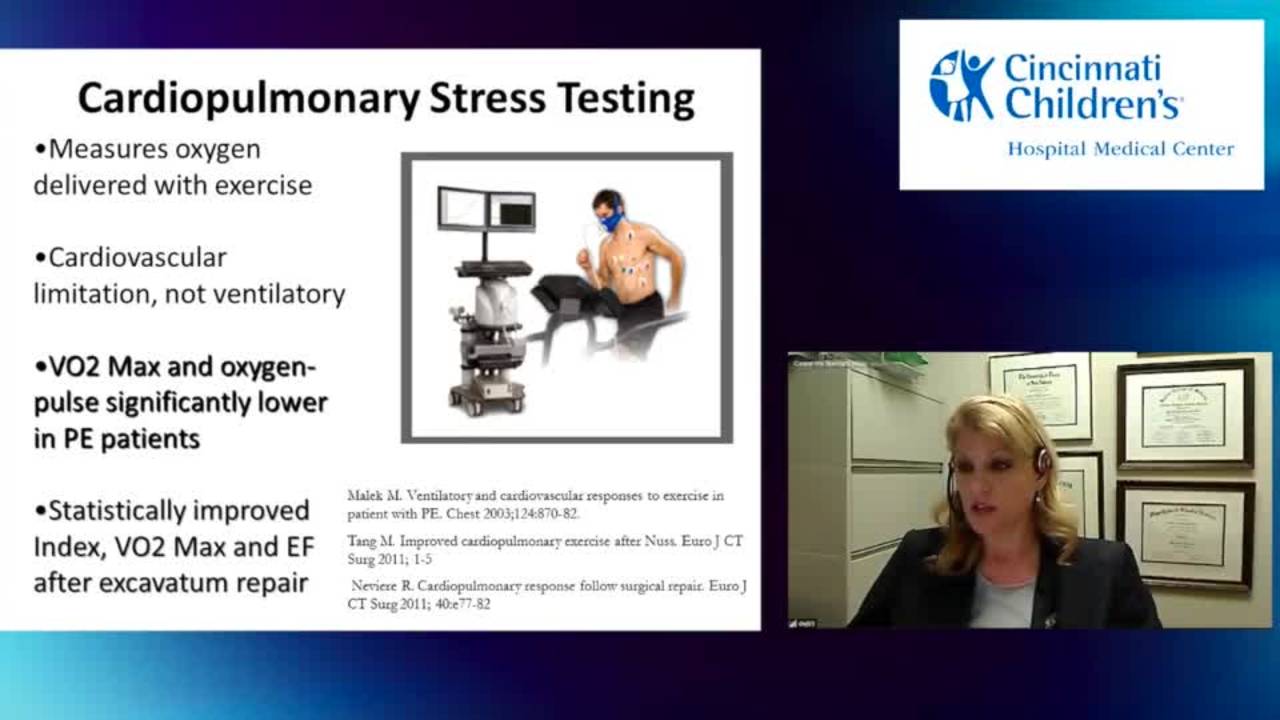
  • 19:22Decreased RV function manifests more clearly during formal cardiopulmonary exercise testing — Michael Taylor
  • 22:15Most pediatric MRI centers can perform cardiac MRI for pectus assessment using fairly basic sequences — Eric Crotty
  • 23:00Children tolerate MRI well, especially with video goggle technology allowing them to watch movies during the procedure — Eric Crotty
  • 23:37Cardiac MRI does not require sedation or contrast for pectus assessment — Eric Crotty
  • 23:53MRI can be performed after pectus bar placement with only minimal localized artifact where bar overlies the heart — Michael Taylor
  • 26:19Insurance companies may deny pectus surgery if only Haller index is provided, requiring additional metrics and cardiac dysfunction evidence — Speaker 4
  • 26:50Bundle of correction index, depression index, and cardiac dysfunction evidence is effective in reversing insurance denials — Speaker 4
  • 27:32When sternum presses on RV free wall, it affects right ventricular function — Michael Taylor
  • 28:49Cardiac MRI reports aortic root dimensions and can detect mitral valve prolapse in Marfan patients — Michael Taylor
  • 21:12Older pectus patients tend to have heart extruded more into left chest, possibly a growth/developmental phenomenon — Michael Taylor
  • 34:13Earlier CT exposure and higher CT dose increase long-term risk of malignancy development — Eric Crotty
  • 34:38Pediatric tissues have more rapidly dividing cells with more time to develop DNA breaks, increasing complication likelihood from CT — Eric Crotty
  • 35:57Even one CT scan shows increased risk of long-term malignancy, with risk increasing the younger the patient — Eric Crotty
  • 36:59Dr. Park in Korea reports that pectus surgery is safe in patients older than 3 years — Becky
  • 37:15Shorter bars are required for younger patients to avoid impeding chest wall growth — Speaker 4
  • 37:45Early correction can convert excavatum to carinatum, particularly in patients with connective tissue disorders — Speaker 4
  • 38:18Dr. Park reports fewer asymmetric pectus cases when correcting patients at younger ages — Speaker 4

Cases discussed

  • 0:1613-year-old male with progressive pectus excavatum, exercise intolerance, and chronic postoperative pain

Points of disagreement

  • 18:34Whether pectus surgery is primarily cosmetic vs medically necessary
    • Speaker 4: Some colleagues view pectus surgery as cosmetic, but cardiac MRI data showing RV dysfunction in 15% of patients and reduced RV ejection fractions supports medical necessity
    • Speaker 3: Expresses initial skepticism, frequently sends patients away saying surgery is cosmetic, has personal pectus and is fine with it, but becoming convinced by the cardiac data presented
  • 29:23Optimal age for pectus surgery
    • Speaker 4: Dr. Park (Korea) argues surgery is safe from age 3 with shorter bars, and earlier correction may prevent asymmetric deformities
    • 50% of audience believes age should be >9-11 years, 30% believe >5 years is acceptable

Open questions

  • If a patient has reduced RVEF at age 8 and is re-scanned at age 15, will RVEF remain stable, worsen to 30%, or normalize?
  • At what age does cardiac extrusion into the left chest become irreversible, and is there a critical window for surgical correction to allow cardiac repositioning?
  • What are the long-term cardiac outcomes in adults with uncorrected pectus excavatum and documented RV dysfunction in childhood?
  • Does earlier surgical correction (age 3-5) truly prevent asymmetric pectus deformities as suggested by Korean experience?
  • What is the optimal age for pectus surgery balancing chest wall compliance, cardiac remodeling potential, and risk of overcorrection?
  • In a young Marfan patient (age 4-6) with high Haller index and exercise intolerance, how much of the exercise limitation is attributable to pectus vs other Marfan manifestations?
This episode was analyzed and enriched by Kai, the Library's AI content creator. Every item links to the moment it comes from — click a timestamp to listen in context.
Written for:

Cardiac MRI for Pectus Assessment: Beyond the Haller Index

The episode's main topic retold as a plain-language walkthrough — what it is, why it matters, and what the speakers concluded. Written by Kai from the episode transcript and reviewed before publishing.

For the care team · Explainer · AI-written, human-reviewed

Why This Exists

Pectus excavatum — the inward depression of the anterior chest wall — sits at the intersection of cosmetic concern and cardiopulmonary pathology. For decades, the Haller index (transverse chest diameter divided by anteroposterior diameter, with >3.25 considered abnormal) served as the primary objective measure for surgical candidacy 7:01. But that threshold was observational, not rigorously derived, and it fails in a substantial subset of patients with clinically obvious deformities who measure normal on imaging 5:21. Insurance denials follow, as do missed opportunities to intervene before functional decline becomes symptomatic. The shift from CT to cardiac MRI represents an attempt to solve both problems at once: eliminate radiation exposure in a young population while capturing the functional cardiac data that CT cannot provide 2:31.

The Core Problem

The chest wall deformity compresses the right ventricle. In approximately 15% of patients, right ventricular ejection fraction falls below 50% — the lower limit of normal 16:18. These children are often asymptomatic at rest 19:15, a phenomenon familiar in congenital heart disease, where compensatory mechanisms mask dysfunction until exercise unmasks it 19:22. The clinical challenge is identifying which patients have true cardiopulmonary impairment rather than an isolated cosmetic concern, and doing so with metrics that satisfy both clinical judgment and insurance requirements 26:19.

The Haller index alone is insufficient. Respiratory phase alters the measurement significantly — some patients with abnormal indices at end expiration normalize at end inspiration 4:47 5:21. Barrel-chested patients can have severe deformities with normal Haller indices because the ratio fails to account for chest shape 9:17. A patient denied coverage based on a single borderline number may in fact have measurable cardiac dysfunction that would justify intervention.

How Cardiac MRI Works in This Context

Cardiac MRI delivers anatomical and functional assessment in a single 25-minute study, without contrast or radiation 11:25. The anatomical component replicates what CT provides: precise measurement of the depression depth, sternal tilt, and three-dimensional geometry of the defect 17:04. But MRI adds two indices that address the Haller index's blind spots. The correction index quantifies what percentage of the sternal depression must be elevated to normalize chest configuration; values above 10% show no overlap between normal and pectus patients 8:22. The depression index accounts for variable chest shape — barrel versus elliptical — and a threshold above 0.2 distinguishes abnormal from normal regardless of configuration 8:46.

The functional component is where MRI diverges from CT. Cine imaging captures right ventricular motion throughout the cardiac cycle, yielding ejection fraction, stroke volume, and chamber dimensions. Echocardiography, the traditional tool for this, produces poor image quality in pectus patients because the chest wall deformity distorts the acoustic window 13:23. MRI visualizes the anterior surface of the right ventricle directly, revealing compression that echocardiography cannot see 14:05. When the sternum presses on the RV free wall rather than the RA-RV groove, right ventricular function is more severely impaired 18:14 27:32. This anatomical detail matters: it predicts which patients have depressed ejection fractions and may benefit most from surgical correction.

MRI also screens for associated pathology common in connective tissue disorders — aortic root dilation, mitral valve prolapse — that influence surgical planning and long-term follow-up 28:49. The study can be performed after bar placement with only minimal localized artifact 23:53, making it useful for postoperative assessment as well.

Where Practice Remains Contested

The clinical significance of reduced RV function in asymptomatic adolescents is not fully resolved. These patients tolerate ejection fractions in the 40s without overt symptoms 19:15, and whether early surgical correction prevents long-term cardiac remodeling or whether the heart compensates adequately into adulthood remains unclear. Formal cardiopulmonary exercise testing may unmask dysfunction that resting imaging does not 19:22, but it is not routinely performed. The natural history of untreated pectus with documented RV impairment is incompletely characterized.

Radiation risk from a single chest CT — approximately 7 millisieverts, equivalent to two years of background exposure 35:09 — must be weighed against the diagnostic yield. Younger patients face higher lifetime malignancy risk from ionizing radiation 34:13 34:38 35:57, but the absolute risk from one study remains low. Whether MRI's superior information justifies its cost and longer acquisition time in every case, or whether it should be reserved for equivocal cases and those requiring cardiac assessment, is a matter of institutional practice and payer policy.

Optimal surgical timing is debated. Most North American surgeons favor repair after age 9 to minimize recurrence and avoid impeding chest wall growth. Korean experience suggests safety and efficacy as young as age 3, using shorter bars to accommodate growth 36:59 37:15. Early correction may reduce the incidence of asymmetric deformities 38:18, but it carries a risk of overcorrection — converting excavatum to carinatum, particularly in patients with connective tissue disorders 37:45.

When to Involve This Team

Refer patients with progressive pectus excavatum and any of the following: exercise intolerance, chest pain, palpitations, or a Haller index approaching 3.25 on CT. Refer earlier if there is clinical suspicion of Marfan syndrome or another connective tissue disorder, as these patients require aortic surveillance regardless of pectus severity. If a patient has been denied insurance coverage based on Haller index alone but you believe the deformity is functionally significant, cardiac MRI with correction index, depression index, and RV function assessment provides the objective data to appeal that denial 26:50. The bundle of metrics — not the Haller index in isolation — is what reverses coverage decisions.

Takeaways from this story

  • 15% of pectus patients have RV ejection fraction <50% despite being asymptomatic at rest; cardiac MRI detects this where echo fails.
  • Haller index normalizes at end inspiration in some patients with clinically obvious deformity; correction and depression indices fill the gap.
  • Insurance denials based on Haller index alone can be reversed with bundled MRI metrics: correction index, depression index, and RV function.
  • A single chest CT delivers 7 mSv — two years of background radiation — with higher lifetime malignancy risk the younger the patient.
  • Compression of the RV free wall (vs RA-RV groove) predicts more severe functional impairment; MRI visualizes this, echo cannot.

Keywords

Hashtags

Transcript

Comments

Loading comments…