
Physiology: Pectus Innovations
Part of
Pectus Excavatum 58 items
Educational content from recorded physician discussions — not medical advice. Talk to your (or your child's) care team about your situation.
Video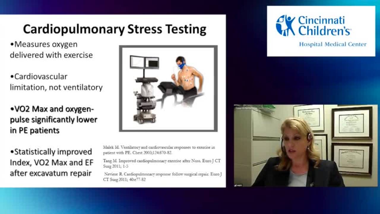
Pectus - Physiologic Consequences and Research
22 min · Published Nov 2018
Video
Pectus - Preoperative Assessment - Radiology and Cardiac Evaluation
Dr. Todd Ponsky · 44 min · Published Nov 2018
Video
Radiology: Pectus Innovations
38 min · Published Oct 2015
Video
Criteria for Pectus Repair: Update Course 2015
55 s · Published Nov 2015
Video
Vacuum Bell Therapy for Pectus Excavatum: Long-term Experience at a Single Center
58 s · Published Apr 2025
Video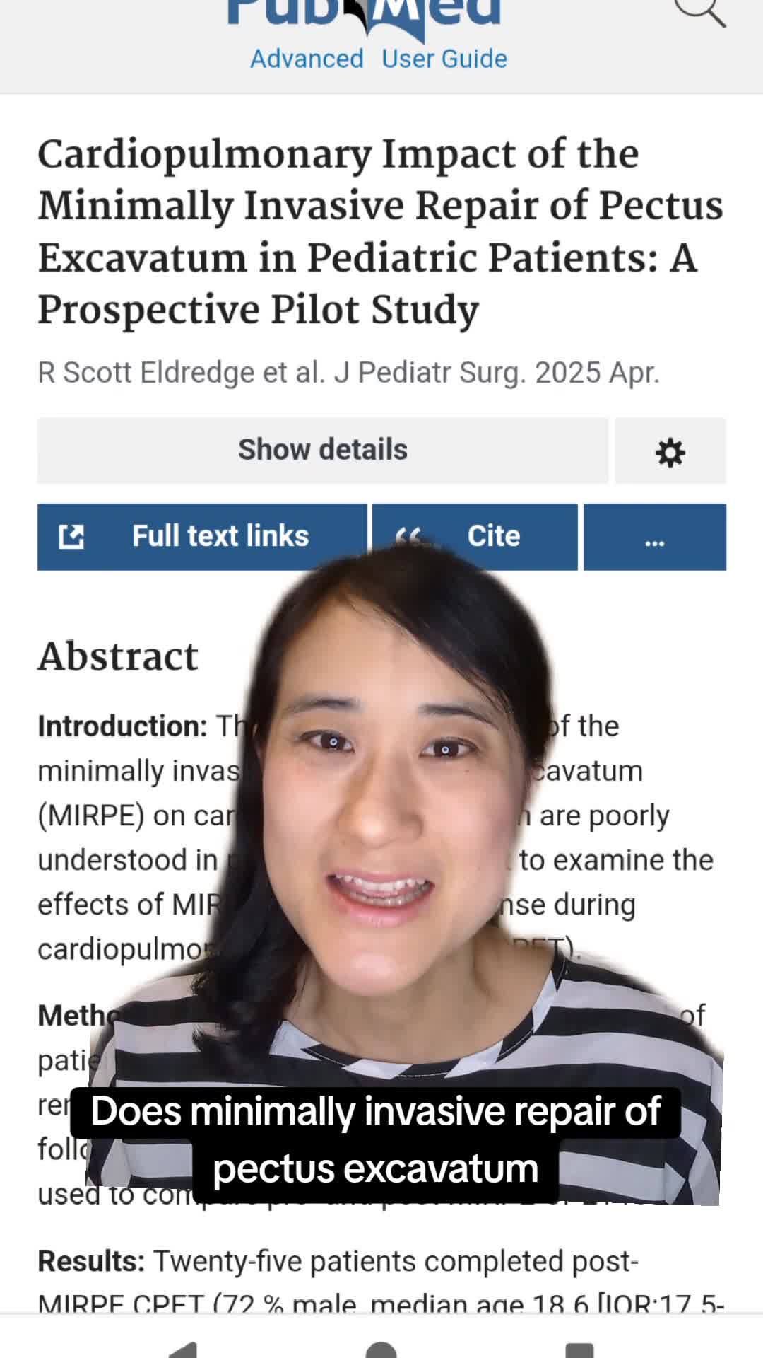
Cardiopulmonary Impact of the Minimally Invasive Repair of Pectus Excavatum in Pediatric Patients: A Prospective Pilot Study
55 s · Published Apr 2025
Video
Pediatric Surgical Oncology Research Collaborative (PSORC): Studying Rare Pediatric Tumors
56 s · Published May 2026
Video
Update Course Rewind 2025: Hirschsprung + ARM: Rare but Real
1 min · Published May 2026
Video
Update Course Rewind 2025: Hirschsprung + ARM: Rare but Real
1 min · Published May 2026
Video
Pooling Patients to Study Rare Pediatric Tumors: An Introduction to PSORC
56 s · Published May 2026
Video
The fetal frontier: A review of current and emerging fetal therapies for genetic diseases
44 s · Published May 2026
Video
Indocyanine green assists with sentinel lymph node mapping in pediatric and adolescent patients
1 min · Published May 2026
What the experts said
Older pectus patients report progressive exercise intolerance and breathing difficulty developing over the last decade after being asymptomatic their whole life.
99% of pectus patients have transthoracic echocardiograms reported as normal with no issues.
Transthoracic echo has difficulty obtaining adequate windows to visualize the right side of the heart in pectus patients.
Intraoperative transesophageal echocardiography shows marked improvement in right ventricular chamber dimensions after pectus repair.
Flow velocity through the right ventricular outflow tract nearly triples after pectus repair.
Left ventricular output significantly improves when right ventricular flow increases after pectus repair because the heart functions as a pump.
Cardiac output can improve from 55% (normal range) to 77% after pectus repair in older patients, with patients noticing improved exercise capacity and reduced fatigue.
Cardiac contractility, synchrony, and mobility show significant differences between pre-operative and post-operative states in pectus patients.
The efficiency of the cardiac pump improves after pectus repair, likely due to correction of heart distortion and right ventricular chamber deflection.
In a series of 168 patients, minimally invasive repair (MIRP) showed 30% increase in cardiac output while open repair showed less improvement, possibly because open cases include mixed pathology like malunion.
Overall cardiac output increased 24% across all pectus repair patients in the series.
Pectus excavatum is a medical disease, not cosmetic, and affects patients physiologically, with worsening tolerance to compression as they age.
Standard Bruce protocol ischemic stress testing is not appropriate for pectus evaluation unless ischemia is suspected; cardiopulmonary exercise testing is needed instead.
Cardiopulmonary exercise testing evaluates VO2 max and anaerobic VO2, which are very abnormal in pectus patients while other parameters remain normal, and separates cardiac from pulmonary components.
A French study of 125 adults showed cardiopulmonary exercise parameters statistically improve and normalize after surgical pectus repair.
Pectus patients show limited ability to increase stroke volume during exercise; output initially increases by heart rate but plateaus when volume increase is needed due to chest cage restriction, appearing as a flat line rather than the normal upward slope.
The flat stroke volume response to exercise improves statistically significantly after pectus repair.
Cardiopulmonary exercise testing is used routinely in congenital heart disease to assess functional capacity and guide decisions about palliation and conduit replacement.
Cardiopulmonary exercise testing has not been performed frequently for pectus patients because surgical decisions have been made on other bases and testing has not been required.
Cardiopulmonary exercise testing would be a good tool for borderline pectus cases where there is uncertainty about proceeding with surgery.
Ehlers-Danlos type 4 (vascular type) does not typically present with pectus excavatum as it is not primarily a skeletal or hypermobile disorder.
Ehlers-Danlos type 4 involves internal tissue fragility requiring specially managed repairs, and would require careful consideration before proceeding with pectus surgery.
Genetic testing should be performed to confirm Ehlers-Danlos type 4, and skin biopsy may be needed if genetic testing is normal but clinical suspicion remains high.
Patients frequently self-diagnose Ehlers-Danlos vascular type based on internet research, visible veins, or family history of abdominal aortic aneurysm, none of which actually indicate the disorder.
There is no genetic disorder that represents an absolute contraindication to pectus surgery, only relative contraindications based on functional status.
Very young children may outgrow their pectus bars and require explantation and replacement, representing a cost of early repair.
Adolescents who are nearly completely grown may have better cost-benefit ratio for pectus repair compared to very young children.
A seven-year-old with severe symptomatic pectus may need earlier repair despite general preference to wait.
An Easter Seals grant study at UCLA using cardiopulmonary exercise testing in children showed that while some classes of patients had deficits, a large percentage of children did not show problems.
Compensation mechanisms work better in younger pectus patients; deficits become more apparent on testing as patients age.
Some children are completely asymptomatic and progress to become symptomatic as they age, while others are symptomatic when very young.
Young children have more pliable chest walls, allowing repair with minimal force (described as 'a popsicle stick'), while adults have tremendous pain due to chest wall rigidity.
In Korea, pectus repairs are performed on 3-4-5 year olds using very small bar sets.
The trend in the United States has shifted from repairing pectus patients young to repairing them at older ages.
The institution where Donald Nuss practices has moved away from performing pectus repairs on young children, suggesting they encountered more issues than reported in initial series, possibly identified during long-term follow-up.
Korean surgeons (specifically Yung Park) routinely repair pectus in young children and report no problems, considering it unusual to wait until older ages.
Vacuum bell therapy works if the patient has a very flexible chest and wears the device religiously.
Patient compliance with vacuum bell therapy varies greatly, with many young children tolerating it for only short periods despite parental efforts.
Vacuum bell therapy is recommended for very young children (e.g., 5 years old) as an alternative to operative repair.
Dawn has selection bias in her practice because she predominantly sees symptomatic pectus patients, while thousands of asymptomatic adults with pectus likely exist.
95% of patients presenting to Dawn's office are seeking surgery.
When discussing testing results with pectus patients, they often reveal subtle symptoms they had not previously recognized or reported, such as reduced exercise capacity compared to peers.
Pectus operations are better tolerated when patients are younger, but older patients require substantial narcotics for pain control.
Donald Nuss initially performed pectus repairs on very young patients, as young as 3 years of age, without high recurrence rates in his early series.