
Collection
Long-gap Esophageal Atresia
Educational content from recorded physician discussions — not medical advice. Always talk to your child's care team about your child's situation.
Content of this collection
Resources
4 items
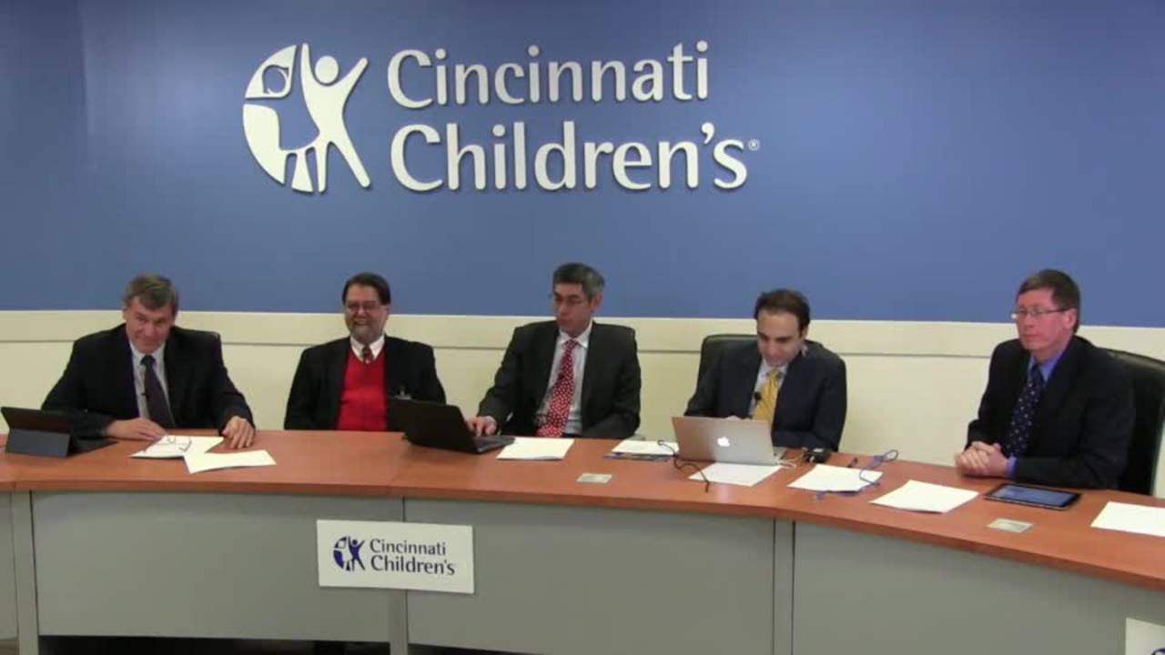


Aerodigestive & Esophageal Surgery - The Unsalvageable Esophagus & Cases
Watch →
Join the multidisciplinary team from Cincinnati Children's Hospital as they review the complexities in the management of Tracheal Esophageal Fistula (TEF). This fast paced live event will feature case presentations, video, and panel discuss
video1:41:09 · Nov 2018
Treatment for Long Gap Esophageal Atresia: Esophageal Elongation and Replacement
Listen →
Pediatric surgeons Dr. Daniel Von Allmen and Dr. Todd Ponsky from Cincinnati children's Hospital discussing the technique for treating long gap esophageal atresia. For additional info please visit: https://www.youtube.com/c/CincinnatiChildr
podcast10:56 · Jan 2022
Journal of Pediatric Surgery Article Review: February 2023, BAPS issue
Listen →
We’re featuring new Journal of Pediatric surgery articles this week to bring you some of the latest news! This week we are discussing three articles from the February 2023 issue, the BAPS issue
We talked to the editor Dr. Mark Davenport
podcast12:32 · Apr 2023
Long Gap Discussion: EA & TEF
Watch →
Dr. Todd Ponsky moderates a panel discussion on the work-up of long gap esophageal atresia.
video10:16 · Jan 2019
Summaries and takeaways+ Show
Summary of this collection+ Show
Key points, with the moment each was said+ Show
Featured picks for families are being prepared — the summaries above are written for you.
Takeaways+ Show
Long-gap esophageal atresia (LGEA) is defined by a gap ≥2 vertebral bodies or ~2 cm, though accurate measurement is challenging—blind probing can push the diaphragm cephalad, overestimating the gap . Primary anastomosis is attempted when feasible; if tension is excessive, placing the ends on internal traction for one week often permits successful delayed closure due to stretch-induced growth [e738-c2, e1077-c15]. Even 2–4 mm esophageal ends will grow into serviceable conduits under traction . The Foker technique (external traction sutures) and van der Zee thoracoscopic variant both exploit this principle [e4791-c2, e4791-c5]. However, these approaches carry significant morbidity: median ICU stays of 70–110 days, prolonged paralysis, and only 10–67% achieving full oral nutrition depending on primary vs. secondary repair [e4791-c8, e4791-c9].
When primary repair is not achievable by 6–8 weeks or the gap exceeds 5–6 vertebrae, esophageal replacement is indicated [e1077-c8, e1077-c9]. Options include gastric pull-up, colonic interposition (full-length or segmental), and small bowel interposition . Segmental colonic interposition preserves the native GE junction, reducing long-term redundancy and permitting future anti-reflux surgery at the native junction [e738-c5, e738-c6]. Full-length colon interposition risks progressive tortuosity and poor drainage [e4791-c14, e738-c47]. Gastric pull-up is recommended as first-line replacement by some groups . Complications are nearly inevitable; the goal is effective anticipation and multidisciplinary management .
- LGEA is defined as gap ≥2 vertebral bodies; accurate measurement requires endoscopy or fluoroscopy to avoid overestimation from diaphragmatic displacement during blind probing.
- Internal traction for one week after initial mobilization permits delayed anastomosis via stretch-induced growth, even from 2–4 mm esophageal ends.
- Foker and van der Zee techniques achieve 96% intact esophagus in de novo cases but require median ICU stays of 70–110 days with prolonged paralysis.
- Esophageal replacement is indicated if primary repair is not achievable by 6–8 weeks or gap exceeds 5–6 vertebrae; options include gastric pull-up and colonic interposition.
- Segmental colonic interposition preserves the native GE junction, reducing long-term redundancy and permitting future anti-reflux surgery, but requires two anastomoses and thoracotomy.
For patients & families
Long-gap esophageal atresia means the two ends of the food pipe are too far apart to connect right away. Doctors measure the gap using special cameras or X-rays with contrast dye [e738-c1, e1077-c4, e1077-c6]. If the gap is about 2 centimeters or less, surgeons can often bring the ends together during an operation . Sometimes they put gentle tension on the ends for a week, which helps them stretch and grow closer together so they can be connected at a second operation [e738-c2, e1077-c15, e4791-c3]. If the gap stays too wide after several weeks of waiting—usually more than 5 or 6 vertebrae by 6 to 8 weeks—doctors may recommend replacing the esophagus with a piece of the child's own colon or stomach [e1077-c8, e1077-c9, e4791-c10, e4791-c11]. These replacement operations are major procedures but can allow children to eat normally over time [e738-c59, e4791-c9]. The medical team watches closely for complications like narrowing (strictures), leaks, or breathing problems, which are common but can be managed [e738-c60, e738-c10]. Every child's situation is unique, and the care team will explain which approach is safest for your baby.
Long-gap esophageal atresia means the two ends of the food pipe are too far apart to connect right away. Doctors measure the gap using special cameras or X-rays with contrast dye [e738-c1, e1077-c4, e1077-c6]. If the gap is about 2 centimeters or less, surgeons can often bring the ends together during an operation . Sometimes they put gentle tension on the ends for a week, which helps them stretch and grow closer together so they can be connected at a second operation [e738-c2, e1077-c15, e4791-c3]. If the gap stays too wide after several weeks of waiting—usually more than 5 or 6 vertebrae by 6 to 8 weeks—doctors may recommend replacing the esophagus with a piece of the child's own colon or stomach [e1077-c8, e1077-c9, e4791-c10, e4791-c11]. These replacement operations are major procedures but can allow children to eat normally over time [e738-c59, e4791-c9]. The medical team watches closely for complications like narrowing (strictures), leaks, or breathing problems, which are common but can be managed [e738-c60, e738-c10]. Every child's situation is unique, and the care team will explain which approach is safest for your baby.
The doctors in this collection+ Show
All expert statements+ Show
Every expert statement below comes from the recorded discussions, with its speaker and moment.
Aerodigestive & Esophageal Surgery - The Unsalvageable Esophagus & Cases
For long-gap esophageal atresia, a gap of 2 vertebral bodies or approximately 2 centimeters is considered close enough to attempt primary anastomosis.
guidelineDan5:44 ↗
If primary anastomosis cannot be achieved at initial operation, placing the ends on tension for one week and then re-operating often allows successful closure due to stretch.
clinicalDan5:51 ↗
In experimental porcine models of endoscopic Foker (using olive beads and wire traction), pressure necrosis at the anastomotic site is the suspected cause of death.
clinicalTodd Ponsky6:32 ↗
Intraluminal magnetic anastomosis is being developed as a minimally invasive approach to long-gap atresia, with the goal of reducing pressure necrosis compared to bead-based traction.
clinicalTodd Ponsky7:00 ↗
Segmental colonic interposition (preserving distal native esophagus) reduces the risk of long-term redundancy and tortuosity compared to full-length colon interposition from cervical esophagus to stomach.
clinicalDan9:00 ↗
Segmental interposition preserves the native gastroesophageal junction, allowing potential future anti-reflux surgery at the normal GE junction rather than at a colonic-gastric anastomosis.
clinicalDan9:22 ↗
Segmental colonic interposition requires two anastomoses (one in chest, one in neck) and a thoracotomy, making it a morbid operation, but it is theoretically 'one and done.'
clinicalDan9:44 ↗
Sometimes surgeons try too hard to salvage the native esophagus, and children may do better with esophageal replacement, especially when chronic aspiration from recurrent TEFs or strictures causes severe pulmonary disease.
opinionDan11:24 ↗
When mobilizing a distal esophageal pouch off tension (e.g., from prevertebral fascia), the pouch retracts and becomes much shorter than it appeared pre-operatively.
clinicalDan12:53 ↗
Esophageal stents can compress the adjacent trachea, especially in children with tracheomalacia or when the esophagus lies directly posterior to the trachea. Always perform bronchoscopy after stent placement.
Host summaryEm Gootee summarizing the discussion — not the host's own clinical position48:18 ↗
On CT scan, if the dilated esophagus lies immediately posterior to the malacic trachea, any esophageal distention (food bolus, stent) will compress the trachea. If the esophagus deviates to the left, tracheal compression is less likely.
clinicalBob48:58 ↗
Chronic esophageal foreign bodies (e.g., coins lodged for weeks) can cause posterior tracheal bulge and present as refractory asthma or bronchiolitis, often diagnosed late because chest X-rays are not routinely obtained in asthma protocols.
Host summaryEm Gootee summarizing the discussion — not the host's own clinical position50:20 ↗
For recalcitrant esophageal strictures, weekly dilations (3–4 cycles) prevent fibroblasts from bridging and allow the stricture to scar open rather than closed.
Host summaryEm Gootee summarizing the discussion — not the host's own clinical position36:29 ↗
Needle knife incision of esophageal strictures is effective for short, well-defined, non-circumferential scar bands. It should be avoided on the anterior wall in patients with prior TEF due to proximity to the trachea.
clinicalPhil35:13 ↗
Kenalog (steroid) injection after stricture dilation is preferred over mitomycin. Mitomycin at high concentrations (up to 5 mg/mL) causes tissue necrosis and has not shown superior outcomes compared to steroids.
Host summaryEm Gootee summarizing the discussion — not the host's own clinical position33:28 ↗
Patients born with tracheoesophageal fistula, especially those with esophageal atresia, are at significantly higher risk for eosinophilic esophagitis, a non-acid inflammatory condition that causes strictures if untreated.
epidemiologicalPhil46:14 ↗
Eosinophilic esophagitis in infants is effectively managed with elemental formula, which works in approximately 95% of cases.
clinicalPhil47:14 ↗
Routine esophageal biopsies should be performed during endoscopy in TEF patients to screen for eosinophilic esophagitis, even in the absence of overt symptoms.
guidelinePhil46:53 ↗
In patients with recalcitrant anastomotic strictures and documented reflux, treating reflux with fundoplication may allow the stricture to heal. However, this approach risks making subsequent esophageal mobilization more difficult if replacement is ultimately needed.
clinicalDan43:07 ↗
Medical acid suppression (PPI) reduces gastric acid but does not stop reflux itself. A mechanical barrier (fundoplication) is required to prevent reflux of bile and other irritants.
clinicalPhil44:13 ↗
Feeding via gastrojejunal tube with gastric drainage (GJ/G) is a temporizing measure to reduce reflux into the esophagus while managing a refractory stricture.
Host summaryEm Gootee summarizing the discussion — not the host's own clinical position45:47 ↗
Substernal colonic interposition avoids thoracotomy and is useful when the chest is heavily scarred from prior operations. However, it requires sacrificing the entire native esophagus.
clinicalDan10:27 ↗
When performing segmental colonic interposition, the choice of right vs. left colon is based on intraoperative assessment of the marginal artery; the side with better vascularity is selected.
clinicalDan1:06:15 ↗
Redundant colonic interposition can be revised by shortening the conduit at the distal anastomosis, dividing small branches along the colonic wall while preserving the vascular arcade.
clinicalDan1:12:54 ↗
Covered esophageal stents (8 mm diameter) are now available for pediatric use and may be effective for temporizing strictures or leaks. They are partially covered to reduce migration and perforation risk.
clinicalPhil38:45 ↗
In complex esophageal cases, a combined operating room evaluation with rigid bronchoscopy, flexible bronchoscopy, and EGD (often with two scopes simultaneously) provides comprehensive anatomic assessment and is safer than sequential procedures.
clinicalDan1:18:10 ↗
Contrast studies through the endoscope under fluoroscopy are routinely performed before attempting wire passage or dilation to confirm anatomy and avoid perforation.
clinicalPhil1:18:53 ↗
Esophageal bypass (leaving native esophagus in situ) is a viable option when resection would create an unreconstructible tracheal defect. A fundoplication is performed to prevent reflux into the residual esophageal pouch.
clinicalDan1:26:54 ↗
When performing esophageal bypass, leaving a tracheoesophageal fistula open allows native esophageal secretions to drain into the airway, preventing mucocele formation in the residual pouch.
clinicalBob1:27:49 ↗
Children who have been unable to swallow for years may continue to spit reflexively even after successful esophageal reconstruction. It can take weeks to months for them to relearn swallowing.
clinicalDan1:28:42 ↗
What's new+ Show