
Prenatal intervention for Fetal Urinary Tract Obstruction
With Dr. Jose Pierro · StayCurrentMD
Cued at 3:29 · stops at 4:14 · press play
Educational content from recorded physician discussions — not medical advice. Talk to your (or your child's) care team about your situation.
Video
Overview of Prenatal Diagnosis: Cincinnati Fetal Center
40 min · Published Nov 2018
Video
Fetal urological aspect: Fetal Genitourinary Disease 2015
59 min · Published Jul 2017
Video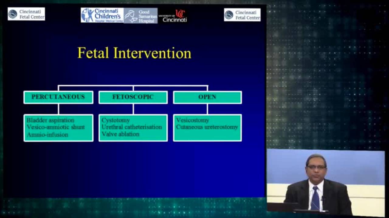
Urologic Fetal Intervention: Cincinnati Fetal Center
61 min · Published Nov 2018
Video
Prenatal diagnosis of fetal lower urinary tract obstruction: Fetal...
40 min · Published Jul 2017
Video
Urologic intervention: Cincinnati Fetal Center
28 min · Published Nov 2018
Video
Fetal management of advanced chronic kidney disease: Fetal Genitourinary...
34 min · Published Jul 2017
Only a few other public items share this expert — go deeper there →
Video
Pediatric Surgical Oncology Research Collaborative (PSORC): Studying Rare Pediatric Tumors
56 s · Published May 2026
Video
Update Course Rewind 2025: Hirschsprung + ARM: Rare but Real
1 min · Published May 2026
Video
Update Course Rewind 2025: Hirschsprung + ARM: Rare but Real
1 min · Published May 2026
Video
Pooling Patients to Study Rare Pediatric Tumors: An Introduction to PSORC
56 s · Published May 2026
Video
The fetal frontier: A review of current and emerging fetal therapies for genetic diseases
44 s · Published May 2026
Video
Indocyanine green assists with sentinel lymph node mapping in pediatric and adolescent patients
1 min · Published May 2026
What the experts said
Prenatal diagnosis of fetal urinary tract obstruction requires ultrasound assessment of keyhole sign, bladder thickness, amniotic fluid volume, and fetal sex.
In male fetuses, posterior urethral valves can be diagnosed and are amenable to fetal intervention.
Fetal urinary tract obstruction causes pulmonary hypoplasia due to anhydramnios (insufficient amniotic fluid leading to underdeveloped lungs).
Obstructive nephropathy from urinary tract obstruction creates renal dysplasia, leading to renal insufficiency with associated morbidity, mortality, and need for transplant.
A distended bladder without cycling contractions produces bladder dysfunction with infections and other problems postnatally.
Therapeutic goals for fetal urinary tract obstruction are to restore amniotic fluid (protect lungs), decompress the urinary system (protect kidneys), and allow detrusor cycling (protect bladder function).
Renal function assessment in fetal urinary obstruction is based on bladder tap (fetal urine analysis), ultrasound evaluation of renal parenchyma for cystic dysplasia, and bladder refilling after bladder tap.
Bladder refilling after vesicocentesis demonstrates that the fetus has the ability to make urine.
In severe renal dysplasia where the fetus makes no urine, the only treatment goal is pulmonary survival by replacing amniotic fluid; kidney function cannot be improved.
In moderate or borderline renal dysplasia where kidneys make small amounts of urine, intervention is likely too late to save kidney function but can save the lungs and provide the option of renal transplant.
In cases with no renal dysplasia where kidneys make plenty of urine, decompressive techniques can theoretically rescue both kidney and lung function.
Vesico-amniotic shunt devices commonly used include Rodeck, Harrison, Cook, and Summaex (more common in Europe).
Vesico-amniotic shunt placement has a 98% technical success rate but a 15% complication rate, with complications carrying 100% mortality due to loss of amniotic fluid.
Overall survival with vesico-amniotic shunting is approximately 50%, and of survivors, 40% will develop end-stage renal disease.
Open fetal surgery for vesicostomy was attempted by Crombleholme and Lim in Cincinnati, using minimal uterine exposure to create an abdominal wall opening and marsupialize the bladder for drainage, allowing spontaneous postnatal closure.
In the Cincinnati Children's Hospital series, 6 male fetuses between 19-22 weeks gestational age with anhydramnios and normal fetal urine electrolytes underwent open vesicostomy; all achieved complete postnatal urinary tract decompression, but only 2 of 6 survived, with no maternal complications.
The main technical challenge with fetal cystoscopy for posterior urethral valves is the angulation; while bladder access is good, visualization and orientation of the posterior urethral valves during surgery can be difficult.
Transurethral catheter placement can be combined with laser therapy for posterior urethral valve treatment; in one case at 23 weeks, a wire was used to identify and access valves, followed by laser ablation and transurethral catheter deployment.
For fetuses with kidney dysplasia and minimal urine output who are not candidates for shunt or cystoscopy, the only option is attempting lung rescue by placing fluid from outside to restore amniotic fluid.
Methods for amniotic fluid restoration include repeated serial amnio-infusions with needle (once weekly initially, twice weekly in late gestation) or placement of an amnio-port.
Amnio-port is a metal port used for intravascular medication and parenteral nutrition that can be implanted inside the uterus for easy subcutaneous access to replace amniotic fluid.
Complications of vesico-amniotic shunting include blockage, shunt migration (common as fetuses pull or dislodge the shunt), preterm delivery, urinary ascites, chorioamnionitis, iatrogenic gastroschisis, and abdominal or inguinal herniation.
A Birmingham University trial comparing percutaneous shunting to expectant management (15 vs 16 cases) showed a trend toward improved perinatal survival with shunting.
A meta-analysis of 9 papers showed perinatal survival advantage with vesico-amniotic shunt, but 1-2 year survival and renal function outcomes were unclear.
Open fetal surgery requires deep general anesthesia, maternal laparotomy and hysterotomy, and carries increased maternal and fetal risk with higher prematurity rates.
Fetal cystoscopy provides endoscopic surgical access to the fetus, amniotic cavity, umbilical cord, and fetal side of the placenta through a small maternal abdominal incision.
Cincinnati Children's Hospital prefers the mini-laparotomy approach for fetal cystoscopy over percutaneous access because it provides better visualization of posterior urethral valves using T-fasteners to align tissue layers.
Fetal cystoscopy can be used therapeutically for valve ablation, with the goal of fulguration to maintain an open urethra.
A study by Ruano et al. found 60% survival with therapeutic fetal cystoscopy compared to 10% in the control group.
A 2015 study by Ruano et al. comparing fetal cystoscopy, VA shunting, and no intervention found survival rates of 66%, 60%, and 17% respectively.
The 2015 Ruano study suggested fetal cystoscopy decreased the incidence of chronic kidney disease in patients with posterior urethral valves.
A 2019 VA study found no difference between VA shunting and fetal cystoscopy regarding chronic kidney disease incidence or survival.
Ureterocele can cause bladder obstruction by prolapsing through the posterior urethra; in females it can prolapse completely out due to the short urethral distance, and can be treated surgically by colon puncture or cystoscopy-assisted laser incision to decompress the cyst.
In the only published paper on amnio-port outcomes, there were no cases of fetal death related to the procedure, complete restoration of amniotic fluid in all cases, and pulmonary hypoplasia at birth in 6 of 8 fetuses.