
The Full Story on CPAMs
With Dr. Dr. Pam Choi and Dr. Beth Romesky & Dr. Dr. Jack Langer & Dr. Dr. Steve Rothenberg · hosted by Dr. Rod Gerardo and Ellen Ancisco · StayCurrentMD
Cued at 13:33 · stops at 14:18 · press play
Educational content from recorded physician discussions — not medical advice. Talk to your (or your child's) care team about your situation.
More about CPAM (congenital pulmonary airway malformation)
same diagnosisVideo
Management of Asymptomatic Lung Lesions: Pediatric Thoracic Surgery Part...
Dr. Todd Ponsky · 20 min · Published Jul 2017
Video
Prenatal Management of CPAMs: Lung Lesions
18 min · Published Aug 2012
Video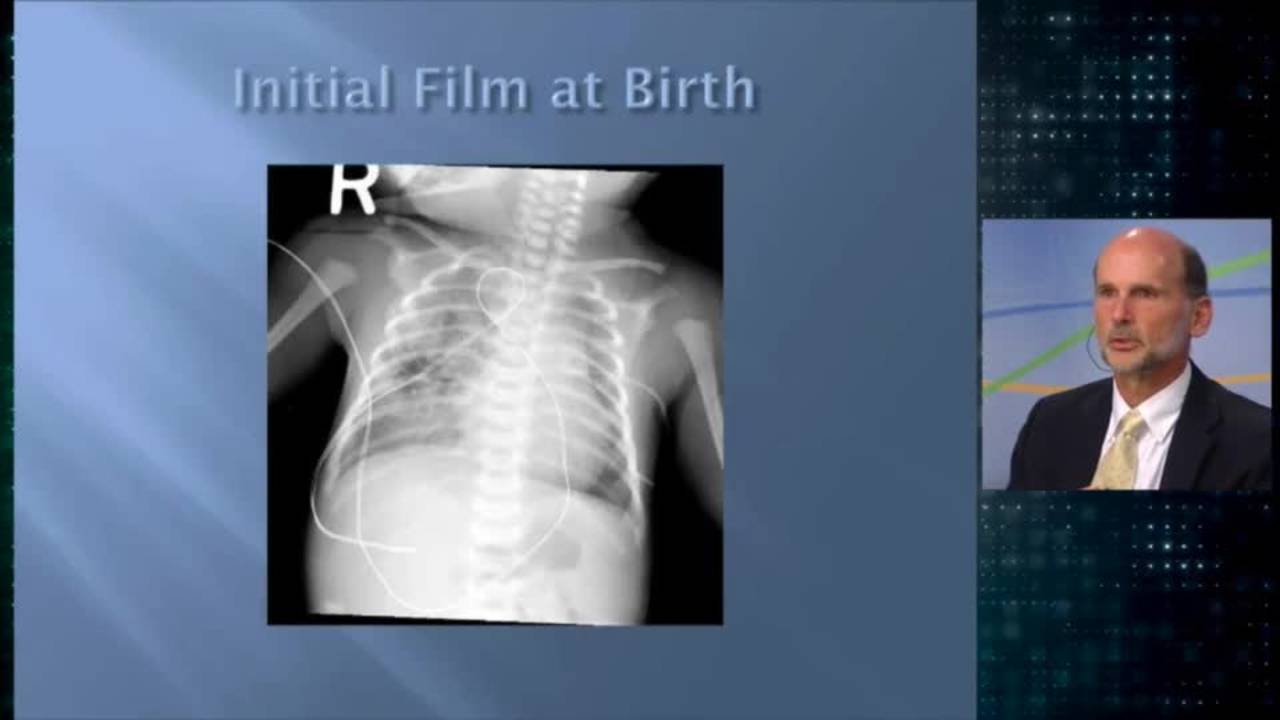
Congenital Cystic Lung Lesions: Update Course 2014
Dr. Steve Rothenberg · 24 min · Published Nov 2018
Video
Open Fetal Surgery & EXIT Procedure with Dr. Jose Peiro
CCHMC Pediatric Surgery · 10 min · Published Oct 2024
More from Dr. Romesky
same expert · first-hand onlyDive deeper → Dr. Dr. Pam Choi and Dr. Beth RomeskyOnly a few other public items share this expert — go deeper there →
Video
Pediatric Surgical Oncology Research Collaborative (PSORC): Studying Rare Pediatric Tumors
56 s · Published May 2026
Video
Update Course Rewind 2025: Hirschsprung + ARM: Rare but Real
1 min · Published May 2026
Video
Update Course Rewind 2025: Hirschsprung + ARM: Rare but Real
1 min · Published May 2026
Video
Pooling Patients to Study Rare Pediatric Tumors: An Introduction to PSORC
56 s · Published May 2026
Video
The fetal frontier: A review of current and emerging fetal therapies for genetic diseases
44 s · Published May 2026
Video
Indocyanine green assists with sentinel lymph node mapping in pediatric and adolescent patients
1 min · Published May 2026
What the experts said
CPAMs are abnormally developed lung tissue that doesn't participate in gas exchange, coming off normal bronchial airways but lacking normal alveoli.
Prenatal steroids (betamethasone 12mg given twice, 24 hours apart) have been shown to resolve hydrops and reduce the size of CPAM lesions such that the CVR can decrease.
A thoracoamniotic shunt is placed so that half is inside the fetal chest and half is in the amniotic space, allowing egress of fluid from the fetal chest into the amniotic space for continued decompression throughout pregnancy.
Fetal resection involves partially delivering the fetus, performing a thoracotomy, removing part of the lung, and placing the fetus back in the uterus to continue the pregnancy.
Mirror syndrome means the mother is showing evidence of what the baby has going on, with the mother developing significant edema, pleural effusion, and other symptoms very quickly.
During an EXIT procedure, the baby remains attached to mom through the umbilical cord while a thoracotomy and resection are performed, with the time available depending on how well mom tolerates it and uterine relaxation, typically ranging from a few minutes to rarely longer than an hour.
Air trapping can occur where air enters the CPAM area as the infant breathes in but doesn't come out as easily, causing progressive hyperinflation of the non-functioning lobe and rapid progressive respiratory distress.
Features suggesting higher risk of PPB include: lesion in more than one lobe, in more than one lung, associated pneumothorax, and DICER1 genetic mutation.
Features suggesting very low risk of PPB are a feeding vessel and prenatal diagnosis.
Dr. Langer follows observed CPAM patients with regular chest X-rays for the first 2 years, expecting to see changes on serial X-rays if a PPB is developing from type 1 to type 2, then follows clinically after that.
If a CPAM becomes infected, it should be treated with IV antibiotics, allowed to settle down, then removed, as infected CPAMs make surgery more difficult with more blood loss and complications.
Single lung ventilation for thoracoscopic lobectomy is obtained by mainstem intubation of the contralateral bronchus, preventing the lung from overinflating during dissection of key vessels or structures.
For thoracoscopic lobectomy, the scope port should be placed over the major fissure in the mid-axillary line, anterior to the tip of the scapula, to allow working from front to back and avoid working in paradox.
When completing an incomplete fissure during lobectomy, work through it layer at a time like finger fracturing during liver lobectomy, starting at the front and working posteriorly until exposing the pulmonary artery.
Vascular control in thoracoscopic lobectomy is everything; dissect out vessels to get enough length to make a seal proximally and distally, then cut partway between seals to check for bleeding before completing division.
The bronchus sits right underneath the pulmonary artery and can be felt to help dissect behind the artery during lobectomy.
When dissecting behind the bronchus during lobectomy, stay hard on the backside of the bronchus and be aware that the pulmonary vein is right behind it.
Do not take the pulmonary vein trunk near the pericardium during lobectomy, because if the device fails, the vessel will retract into the pericardium and the child will bleed to death before you can do anything; ensure enough length away from pericardium for proximal control.
The Stocker classification for CPAMs ranges from type 0 to type 4, moving from proximal to distal in the bronchial tree, with type 1 (distal bronchi/proximal bronchioles) being most common at 60-70% of cases.
CPAMs typically grow during the first 20-25 weeks of gestation and plateau around week 28.
The presence of hydrops is the strongest prognostic indicator of mortality in fetuses with CPAMs.
The CPAM volume ratio (CVR) is calculated as the CPAM's length times width times height times 0.52 divided by the head circumference.
In patients with CVR over 1.6, it is estimated that 75% develop hydrops.
CPAMs are differentiated as microcystic (less than 5 millimeters) or macrocystic (larger than 5 millimeters).
CPAMs are not well-visualized on chest X-rays and can regress or get smaller over time.
In the United States, most surgeons resect CPAMs even if asymptomatic, while in Europe and Canada, surgeons sometimes choose to monitor asymptomatic lesions and avoid surgery.
The main reasons for operating on asymptomatic CPAMs are: risk of malignancy both at resection and from malignant transformation later, risk of infection that will make surgery more difficult, and better compensatory lung growth if operating sooner in life.
Thoracoscopic lobectomy risks include bleeding, prolonged post-op mechanical ventilation, infections, air leak, pneumothorax, nerve injury, and mortality.
The particular malignancy associated with CPAM is pleuropulmonary blastoma (PPB), with risk that the lesion at birth already has malignancy and risk of malignant transformation over a person's life.
A recent study published in April 2021 found that no prenatally diagnosed lesions had malignancy, but 10% of patients diagnosed postnatally had malignancy in their resected mass.
The incidence of CPAMs is estimated to be about 1 in every 8,000 to 35,000 births.
60% of prenatally diagnosed lung lesions are CPAMs, making them the most common prenatal lung lesion.