
Collection
Congenital Pulmonary Airway Malformation
Educational content from recorded physician discussions — not medical advice. Always talk to your child's care team about your child's situation.
Content of this collection
Resources
24 items
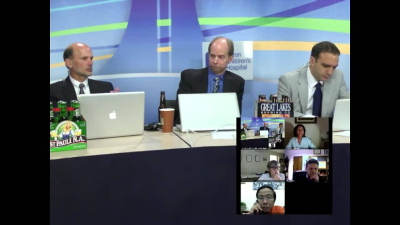


Panel Discussion: Pediatric Thoracic Surgery Part 1-Lung Lesions 2012
Watch →
The internationalfaculty debate and discuss management of difficult cases, includinglower lobe cystic lesion, spontaneous pneumothorax and unilateral bleb disease, and right pulmonary cyst.
video1:01:56 · Jan 2019
Open Fetal Surgery & EXIT Procedure with Dr. Jose Peiro
Watch →
Join us for an insightful discussion with Dr. Jose Peiro, pediatric surgeon and director of endoscopic fetal surgery at Cincinnati Children’s Fetal Care Center, which is the highest-volume fetoscopic center in the nation. Dr. Peiro walks us
video10:23 · Oct 2024
Prenatal Management of CPAMs: Lung Lesions
Watch →
Dr. Alan Flake discusses prenatal management of congenital pulmonary airway malformations (CPAMs). It includes two videos of examples of fetal surgery.
video18:16 · Jan 2019
Should We Resect Asymptomatic CPAM Flake vs Langer
Watch →
Great Debate from 2017! Watch Alan Flake and Steve Rothenberg debate Jack Langer and Jean Matin Laberge on wether or not asymptomatic CPAM's need to be resected. Originally aired March 23, 2017
video14:03 · May 2019
Neonatal Lung Lesions with Dr. Steven Rothenberg
Listen →
Dr. Steven Rothenberg discusses Newborn Lung Lesions with Dr Todd Ponsky. Edited by Ian C. Glenn, MD & Nicholas E. Bruns, MDPrenatal evaluationPatients with prenatal diagnosis of cystic lung lesions are referred for prenatal evaluation and
podcast1:06:58 · Dec 2020
Postnatal Management of Lung Lesions Part III: Pediatric Thoracic Surgery...
Watch →
Course directors Drs Alan Flake, Steve Rothenberg and Todd Ponsky, along with additional faculty including Drs Jean Martin Laberge, Ramesh Santhanakrishnan, Giovanna Riccipetitoni, Mark Wulkan, Alexander Razumovskiy, Starla Martinez, Marcel
video27:56 · Sep 2018
Postnatal Management of Lung Lesions Part I: Pediatric Thoracic Surgery Part...
Watch →
Course directors Drs Alan Flake, Steve Rothenberg and Todd Ponsky, along with additional faculty including Drs Jean Martin Laberge, Ramesh Santhanakrishnan, Giovanna Riccipetitoni, Mark Wulkan, Alexander Razumovskiy, Starla Martinez, Marcel
video31:07 · Sep 2018
Congenital Cystic Lung Lesions: Update Course 2014
Watch →
Dr. Steve Rothenberg, Chief of Pediatric Surgery at Rocky Mountain Hospital for Children, Denver, Colorado, presents the topic of congenital cystic lung lesions. His discussion includes various case presentations and comprehensive panel dis
video24:39 · Nov 2018
Management of Asymptomatic Lung Lesions: Pediatric Thoracic Surgery Part...
Watch →
Course directors Drs Alan Flake, Steve Rothenberg and Todd Ponsky, along with additional faculty including Drs Jean Martin Laberge, Ramesh Santhanakrishnan, Giovanna Riccipetitoni, Mark Wulkan, Alexander Razumovskiy, Starla Martinez, Marcel
video20:11 · Jan 2019
Fetal Interventions Part II: Lung Lesions
Watch →
Dr. Alan Flake leads a discussion in fetal interventions for prenatally-diagnosed lung lesions.
video16:30 · Jan 2019
Neonatal Lung Lesions with Dr. Steven Rothenberg
Listen →
Dr. Steven Rothenberg discusses Newborn Lung Lesions with Dr Todd Ponsky. Edited by Ian C. Glenn, MD Nicholas E. Bruns, MD
podcast1:06:58 · Jan 2019
Open Fetal Surgery Overview: Fetal Surgery 2012
Watch →
Dr. Flake, attending surgeon and director of the General Surgery Fellowship Program at The Children's Hospital of Philadelphia,presents a brief overview of open fetal surgery. Topics discussed include the prenatal diagnosis of an abnormal f
video37:55 · Jan 2019
Fetal Interventions Part I: Lung Lesions
Watch →
Dr. Alan Flake leads a discussion in fetal interventions for prenatally-diagnosed lung lesions.
video14:27 · Jan 2019
Lung Lesions: Fetal Interventions Parts I+II
Watch →
Dr. Alan Flake leads a discussion in fetal interventions for prenatally-diagnosed lung lesions.
video30:56 · Jan 2019
Thoracoscopic Upper Lobectomies for Symptomatic Congenital Pulmonary Airway...
Watch →
This video appears in a new pediatric surgery textbook, Clinical Pediatric Surgery: A Case-Based Interactive Approach," by Dr. Sherif Emil. The book is an innovative educational resource that focuses on judgment and decision-making in pedia
video5:58 · Feb 2020
Thoracoscopic Left Lower Lobectomy for Congenital Pulmonary Airway Malformation
Watch →
This video appears in a new pediatric surgery textbook, Clinical Pediatric Surgery: A Case-Based Interactive Approach," by Dr. Sherif Emil. The book is an innovative educational resource that focuses on judgment and decision-making in pedia
video9:59 · Feb 2020
Thoracoscopic Upper Lobectomies for Symptomatic Congenital Pulmonary Airway...
Watch →
This video appears in a new pediatric surgery textbook, Clinical Pediatric Surgery: A Case-Based Interactive Approach," by Dr. Sherif Emil. The book is an innovative educational resource that focuses on judgment and decision-making in pedia
video5:58 · Feb 2020
Thoracoscopic Left Lower Lobectomy for Congenital Pulmonary Airway Malformation
Watch →
This video appears in a new pediatric surgery textbook, Clinical Pediatric Surgery: A Case-Based Interactive Approach," by Dr. Sherif Emil. The book is an innovative educational resource that focuses on judgment and decision-making in pedia
video9:59 · Feb 2020
The Full Story on CPAMs
Listen →
Here we cover Congenital Pulmonary Airway Malformations (CPAMs) through a story with the help of Dr. Todd Ponsky, Dr. Pam Choi, Dr. Beth Rymeski, Dr. Jacob Langer, and Dr. Steven Rothenberg. Hosts: Ellen Encisco & Rod GerardoChapter 1: Pren
podcast56:08 · Sep 2021
Case Based Journal Review - CPAM in 2022
Listen →
Dr. José Campos is back, this time helping us review some of the latest literature on the diagnosis and management of CPAM in children. In this podcast, we're reviewing a typical case with Dr. Todd Ponsky and incorporating literature from t
podcast12:23 · Apr 2022
Clinical Symptoms Affect Treatment and Prognosis in Pediatric Patients with Congenital Pulmonary Airway Malformation
Watch →
New article you should know by Dr. Cecilia Gigena
"Clinical Symptoms Affect Treatment and Prognosis in Pediatric Patients with Congenital Pulmonary Airway Malformation: A Propensity Score Matching Retrospective Cohort Study"
Authors: Guow
video0:55 · Dec 2023
Complications and Beyond
Watch →
This clip from the 2020 Pediatric Surgery Update Course features an entertaining debate from the full panel surrounding unique and complicated cases presented by Jason Fraser, MD; and Miguel Guelfand, MD.
Highlighted Topics Include:
- M
video1:06:36 · Sep 2020
Thoracoscopic Left Lower Lobectomy Technique by Dr. Steven Rothenberg: How I...
Watch →
Dr. Steven Rothenberg from Rocky Mountain Hospital for Children describes his technique for performing a thoracoscopic left lower lobectomy.
video18:52 · Jan 2019
Thoracoscopic resection of intradiaphragmatic bronchopulmonary sequestration
Watch →
This video, presented at IPEG 2019 in Santiago, Chile by Alexander Gibbons, demonstrates a thoracoscopic resection of an intradiaphragmatic bronchopulmonary sequestration. This congenital anomaly is exceedingly rare, with only 17 previously
video3:59 · Mar 2019
Summaries and takeaways+ Show
Summary of this collection+ Show
Key points, with the moment each was said+ Show
Featured picks for families are being prepared — the summaries above are written for you.
Takeaways+ Show
Congenital pulmonary airway malformation (CPAM) represents a spectrum of abnormally developed lung tissue arising from normal bronchial airways but lacking functional alveoli. The Stocker classification (types 0–4) correlates with bronchial tree location; type 1 (distal bronchi/proximal bronchioles) accounts for 60–70% of cases. Prenatal management hinges on the CPAM volume ratio (CVR = length×width×height×0.52/head circumference): CVR >1.6 predicts 75% hydrops risk, the strongest mortality indicator.[e4463-c5,e4463-c6] Maternal betamethasone (12 mg IM twice, 24 hours apart) is first-line therapy for high-risk lesions, resolving hydrops and reducing CVR in >50% of cases.[e4463-c7,e9333-c12] Refractory hydrops may require thoracoamniotic shunt (macrocystic) or open fetal lobectomy (microcystic <30 weeks).[e4463-c9,e9333-c13] Postnatally, most infants are asymptomatic; air trapping can cause rapid respiratory decompensation.[e4463-c13,e5319-c1] A 2021 multicenter study found 0% malignancy in 344 prenatally diagnosed lesions versus 8.7% in 177 postnatally diagnosed masses, suggesting distinct disease entities.[e5319-c8,e5319-c9] Pleuropulmonary blastoma (PPB) remains the primary malignancy concern; features suggesting higher PPB risk include multilobe/bilateral disease, pneumothorax, and DICER1 mutation, while prenatal diagnosis and feeding vessels confer very low risk.[e4463-c19,e4463-c20] Infection risk is estimated at 20–40% lifetime, complicating subsequent resection.[e307-c13,e643-c9] Thoracoscopic lobectomy at 3 months optimizes tissue planes and minimizes inflammation; operating earlier (1–3 months) yields shorter operative times (115 vs 163 minutes at 6–12 months) without increased complications in experienced hands.[e5319-c24,e5319-c26] Vascular control is paramount: dual-seal technique (two seals 4–5 mm apart, partial division to confirm hemostasis) prevents catastrophic hemorrhage; never take the pulmonary vein near the pericardium.[e307-c32,e4463-c30] The prenatal-versus-postnatal diagnosis dichotomy drives current management controversy: North American practice favors elective resection for all CPAMs, while European/Canadian centers increasingly observe prenatally diagnosed asymptomatic lesions given their negligible malignancy risk and low symptom development (0% symptomatic in one 10-year prospective cohort).[e5319-c3,e5319-c11]
- CVR >1.6 predicts 75% hydrops risk; maternal betamethasone rescues >50% of cases, reducing need for fetal surgery to near-zero in most centers.
- Prenatally diagnosed CPAMs have 0% malignancy versus 8.7% in postnatal diagnoses, representing distinct populations; PPB risk is negligible with prenatal diagnosis and feeding vessel.
- Thoracoscopic lobectomy at 3 months optimizes tissue planes (115-min operative time) versus 6–12 months (163 min); dual-seal vascular technique is mandatory to prevent fatal hemorrhage.
- Never ligate pulmonary vein near pericardium—retraction after device failure causes exsanguination before rescue is possible. Ensure 2+ cm length for proximal control.
- North America resects all CPAMs; Europe/Canada observe prenatally diagnosed asymptomatic lesions (0% became symptomatic in 10-year prospective study). Infection complicates 20–40% if observed.
For patients & families
Congenital Pulmonary Airway Malformation (CPAM) is a lung development difference where some lung tissue doesn't work normally for breathing . Most babies with CPAM diagnosed before birth are born healthy and breathing normally [e5319-c1, e307-c10]. Doctors follow these babies with ultrasounds during pregnancy, usually every few weeks [e307-c1, e948-c1]. The size of the CPAM compared to the baby's head (called CVR) helps doctors predict if problems might develop [e1089-c4, e1089-c5]. If the CVR stays below 1.6, serious complications are very rare—less than 5% . Most CPAMs grow until about 25-28 weeks of pregnancy, then stop growing or even get smaller [e1089-c6, e307-c3]. Very few babies need treatment before birth; doctors at major centers might do fetal surgery less than once every few years [e307-c4, e1088-c23]. If the CPAM causes the baby's heart to struggle (called hydrops), doctors may give the mother steroid shots, which help in more than half of cases [e1027-c22, e1089-c12]. For large fluid-filled CPAMs causing problems, doctors can place a tiny drain before birth [e1089-c25, e1089-c26]. After birth, if your baby is breathing well, you'll likely go home and return for a CT scan around 4-6 weeks to see the CPAM more clearly [e307-c10, e2993-c3].
Congenital Pulmonary Airway Malformation (CPAM) is a lung development difference where some lung tissue doesn't work normally for breathing . Most babies with CPAM diagnosed before birth are born healthy and breathing normally [e5319-c1, e307-c10]. Doctors follow these babies with ultrasounds during pregnancy, usually every few weeks [e307-c1, e948-c1]. The size of the CPAM compared to the baby's head (called CVR) helps doctors predict if problems might develop [e1089-c4, e1089-c5]. If the CVR stays below 1.6, serious complications are very rare—less than 5% . Most CPAMs grow until about 25-28 weeks of pregnancy, then stop growing or even get smaller [e1089-c6, e307-c3]. Very few babies need treatment before birth; doctors at major centers might do fetal surgery less than once every few years [e307-c4, e1088-c23]. If the CPAM causes the baby's heart to struggle (called hydrops), doctors may give the mother steroid shots, which help in more than half of cases [e1027-c22, e1089-c12]. For large fluid-filled CPAMs causing problems, doctors can place a tiny drain before birth [e1089-c25, e1089-c26]. After birth, if your baby is breathing well, you'll likely go home and return for a CT scan around 4-6 weeks to see the CPAM more clearly [e307-c10, e2993-c3].
The doctors in this collection+ Show
All expert statements+ Show
Every expert statement below comes from the recorded discussions, with its speaker and moment.
Thoracoscopic Left Lower Lobectomy for Congenital Pulmonary Airway Malformation
The malformation was a hybrid congenital pulmonary airway malformation diagnosed prenatally.
clinical0:02 ↗
The patient was asymptomatic at birth and remained asymptomatic through 9 months of age.
clinical0:11 ↗
CT scan at 4 months showed a left lower lobe CPAM with systemic blood supply arising from the sub-diaphragmatic aorta.
clinical0:17 ↗
The operation was performed at 9 months of age.
clinical0:29 ↗
The left lung is isolated by right main stem intubation.
clinical0:36 ↗
The patient is placed in the right lateral decubitus position with surgeon and assistant operating facing the patient.
clinical0:41 ↗
A large arterial vessel coming through the diaphragm medial to the inferior pulmonary ligament and entering the left lower lobe confirms a hybrid lesion.
clinical0:48 ↗
The inferior pulmonary ligament is divided all the way to the border of the inferior pulmonary vein to mobilize the left lower lobe off the diaphragm.
clinical0:59 ↗
Division of the inferior pulmonary ligament allows stretching of the systemic arterial vessel for skeletonization with hook cautery.
clinical1:11 ↗
The systemic arterial vessel is double-clipped proximally and divided distally with a ligature device.
clinical1:20 ↗
The approach of proximal clipping and distal ligature division is used repeatedly for control of several major vessels.
clinical1:46 ↗
The fissure is completed using ligature and sharp dissection.
clinical1:59 ↗
Division of pulmonary parenchyma in the fissure allows visualization of pulmonary artery branches to the lower lobe.
clinical2:24 ↗
The dissection should be kept as bloodless as possible, with any bleeding from divided parenchyma controlled early with ligature.
opinion2:50 ↗
Excellent visualization is essential for adequate vascular control.
opinion3:13 ↗
A right angle dissector is extremely useful in skeletonizing vessels and gaining adequate distance for safe ligation.
opinion3:41 ↗
Dissection in the fissure always proceeds from medial to lateral.
clinical4:09 ↗
While ligature can be used as the sole method of vessel control, the surgeon prefers to apply clips proximally if adequate vessel length has been achieved.
opinion4:54 ↗
Once the pulmonary artery and all pulmonary parenchyma in the fissure is divided, the bronchus comes into view.
clinical6:21 ↗
The inferior pulmonary vein is skeletonized using a right angle dissector.
clinical6:38 ↗
The junction of the inferior pulmonary vein and the left atrium can be clearly seen after adequate dissection.
clinical7:58 ↗
It is quite common for the inferior pulmonary vein to consist of two major tributaries that join as they approach the left atrium, in addition to one or more small tributaries.
clinical8:15 ↗
When the inferior pulmonary vein has two major tributaries, it is best to dissect each tributary separately.
opinion8:31 ↗
Each pulmonary vein tributary is controlled by clipping on the cardiac side and applying ligature on the pulmonary side.
clinical8:36 ↗
Hook cautery is used to clean the bronchus of surrounding lymphatic and adventitial tissue.
clinical9:19 ↗
An endo-GIA device is introduced directly through the chest wall and used to staple and divide the bronchus.
clinical9:26 ↗
The specimen is extracted by slightly enlarging the posteriormost port site.
clinical9:36 ↗
A chest tube is placed at the end of the operation.
clinical9:41 ↗
The patient was extubated at the end of the procedure.
clinical9:46 ↗
The patient was discharged on the 2nd postoperative day with excellent recovery.
clinical9:46 ↗
What's new+ Show