14 views 0 likes

StayCurrentMD

GCMD Space · View profile →

Thoracoscopic Upper Lobectomies for Symptomatic Congenital Pulmonary Airway...

Video Published 2020-02-24 Updated 2026-08-01

Timestops (3)

Topic Overview

This surgical demonstration presents thoracoscopic upper lobectomy technique in two pediatric patients with symptomatic congenital pulmonary airway malformations following pneumonia. The presenter details the technical approach to left and right upper lobectomies, emphasizing the challenges posed by post-infectious inflammatory adhesions and the systematic dissection sequence: fissure completion, segmental arterial control, venous division, and bronchial stapling. Both cases achieved complete lung re-expansion postoperatively.

Key Takeaways

  • Upper lobectomies are more technically challenging than lower, especially post-infection due to inflammatory adhesions. (0:01)
  • Systematic dissection sequence: complete fissure anteroposteriorly, control segmental arteries, divide vein, staple bronchus. (1:04)
  • Delay superior pulmonary vein division initially to allow cephalad retraction and better exposure of hilar structures. (1:37)
  • Timing matters: resection performed 3 months after pneumonia resolution to reduce but not eliminate inflammatory adhesions. (3:32)
  • Both patients achieved complete lung re-expansion postoperatively despite challenging inflammatory conditions. (5:46)

Inside this episode

Kai, the Library's AI content creator, listened to this episode and mapped who's speaking, the chapters, key claims, and cases. Every item links to the exact moment in the recording.

AI-enriched

Who's speaking

  • Speaker 1 — guest

Chapters

  • 0:01Introduction and Technical Context — Overview of upper lobectomy challenges and video objectives
  • 0:16Case 1: Left Upper Lobectomy — 12-year-old with left upper lobe cystic malformation; complete surgical technique from positioning through inferior pulmonary ligament division
  • 3:27Case 2: Right Upper Lobectomy — 1-year-old with right upper lobe lesion; surgical approach emphasizing differences in younger patient anatomy and vessel size
  • 5:46Outcomes — Postoperative results showing complete lung expansion bilaterally

Key claims

  • 0:01Pulmonary upper lobectomies are more technically challenging than lower lobectomies, especially following infection — Speaker 1
  • 0:39A dual lumen endotracheal tube and low flow chest insufflation were used to collapse the left lung — Speaker 1
  • 0:45Inflammatory adhesions can be seen between the left upper lobe and the chest wall — Speaker 1
  • 1:04The fissure is completed by dividing the pulmonary tissue starting anteriorly and proceeding posteriorly — Speaker 1
  • 1:37The superior pulmonary vein is identified but not divided initially to allow further cephalad retraction of the upper lobe — Speaker 1
  • 1:51A combination of clips and ligature is used to divide the segmental arteries — Speaker 1
  • 2:02Enlarged lymph nodes can be seen in the fissure — Speaker 1
  • 2:06Where distance allows, the segmental vessels are clipped proximally prior to division with the ligature — Speaker 1
  • 2:3410 millimeter clips are used to control each venous tributary separately — Speaker 1
  • 3:00The segmental bronchus to the lingular segments is divided with an endo GIA — Speaker 1
  • 3:20The inferior pulmonary ligament is divided to allow the lower lobe to rise in the chest — Speaker 1
  • 3:32Resection was performed 3 months after resolution of the pneumonia — Speaker 1
  • 3:37Left main stem intubation was used to isolate the right lung — Speaker 1
  • 3:42Segmental inflammatory adhesions are present and have to be taken down and freed before dissection of the upper lobe — Speaker 1
  • 3:51Persistent inflammation of the upper lobe and adhesions to the middle lobe are visible — Speaker 1
  • 4:16The arteries are much smaller in this younger child and can be taken with a ligature after deliberate dissection — Speaker 1
  • 4:30The most superior and largest of the segmental arteries is clipped proximally prior to application of the ligature — Speaker 1
  • 4:43The superior pulmonary vein is in a slightly more superficial plane than the arteries — Speaker 1
  • 5:30The recurrent segmental artery to the upper lobe arising from the main pulmonary trunk is found in the fissure — Speaker 1
  • 5:46Both patients had excellent outcomes with complete expansion of the operated lung — Speaker 1

Cases discussed

  • 0:1612-year-old boy with left upper lobe congenital pulmonary airway malformation
  • 3:271-year-old girl with right upper lobe congenital pulmonary airway malformation
This episode was analyzed and enriched by Kai, the Library's AI content creator. Every item links to the moment it comes from — click a timestamp to listen in context.
Written for:

Thoracoscopic Upper Lobectomy After Pneumonia: Managing Post-Infectious Adhesions in Pediatric Congenital Lung Cysts

The patient case from this episode, retold from presentation to outcome with the decisions made along the way. Written by Kai from the episode transcript and reviewed before publishing.

For the care team · Case narrative · AI-written, human-reviewed

The presentation

A 12-year-old boy presented with left upper lobe pneumonia surrounding a cystic malformation 0:01. Imaging six weeks later showed resolution of the infection but persistence of the cyst [case1]. The decision was made to proceed with resection rather than continued observation — a choice shaped by the technical reality that pulmonary upper lobectomies are more technically challenging than lower lobectomies, especially following infection 0:01. The inflammatory process had already complicated the anatomy; waiting longer would not improve it.

The operative approach

The case was performed thoracoscopically in right lateral decubitus position using four ports, with a dual lumen endotracheal tube and low flow chest insufflation to collapse the left lung 0:39. The first challenge appeared immediately: inflammatory adhesions between the left upper lobe and the chest wall 0:45. These had to be taken down before any hilar dissection could begin.

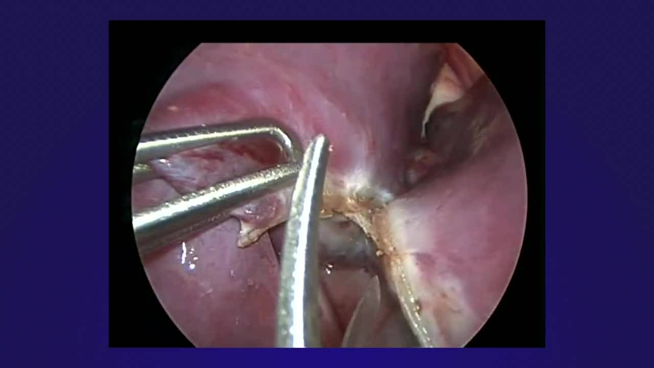
The discussant's sequence is worth noting. Rather than proceeding directly to the hilum, the fissure was completed first — dividing pulmonary tissue from anterior to posterior 1:04. Only then did dissection turn to the vessels. The superior pulmonary vein was identified but deliberately not divided initially, allowing further cephalad retraction of the upper lobe 1:37. This created the working room to identify and dissect the segmental pulmonary arteries.

Vessel control used a combination of clips and ligature 1:51. Where distance allowed, segmental vessels were clipped proximally before division with ligature 2:06. Enlarged lymph nodes were visible in the fissure 2:02 — evidence of the preceding infection. The superior pulmonary vein tributaries were controlled separately with 10 millimeter clips 2:34. The segmental bronchus to the lingular segments was divided with an endo GIA stapler 3:00. Finally, the inferior pulmonary ligament was divided to allow the lower lobe to rise in the chest 3:20.

The second case

A 1-year-old girl presented with a similar picture in the right upper lobe [case2]. Resection was performed three months after resolution of the pneumonia 3:32 — a longer interval than in the first case, though the discussant does not explain why. Left main stem intubation was used to isolate the right lung 3:37.

The same inflammatory picture was present: segmental adhesions that had to be taken down before upper lobe dissection could begin 3:42, and persistent inflammation with adhesions to the middle lobe 3:51. The hilar pleura was divided with hook cautery. The arteries were much smaller in this younger child and could be taken with ligature after deliberate dissection 4:16. The most superior and largest segmental artery was clipped proximally before ligature application 4:30. The superior pulmonary vein was found in a slightly more superficial plane than the arteries 4:43.

In the fissure, a recurrent segmental artery to the upper lobe arising from the main pulmonary trunk was identified and divided 5:30 — an anatomic variant that would have caused troublesome bleeding if missed. The bronchus was divided with an endo GIA.

Outcome

Both patients had excellent outcomes with complete expansion of the operated lung 5:46. The postoperative chest radiographs confirmed full re-expansion on the operative side [case1, case2].

What the case changes

The transferable judgment here is about timing and sequence. These resections were performed weeks to months after pneumonia resolution, not during active infection, but the inflammatory adhesions were still extensive. The discussant's approach — completing the fissure before hilar dissection, leaving the superior pulmonary vein intact initially to improve retraction, clipping larger vessels proximally before ligature division — reflects adaptations to post-infectious anatomy. The alternative would have been to wait longer for inflammation to resolve further, but the discussant's choice implies that the adhesions would not meaningfully improve, and the symptomatic cysts warranted definitive treatment. In a younger child with smaller vessels, the same principles applied but the technical execution shifted toward ligature control rather than clips. The recurrent segmental artery in the second case underscores the need for complete fissure dissection rather than assuming standard anatomy.

Takeaways from this story

  • Upper lobectomies after pneumonia require fissure completion before hilar dissection to manage inflammatory adhesions effectively.
  • Leaving the superior pulmonary vein intact initially allows better cephalad retraction and safer arterial dissection.
  • In younger children, smaller segmental arteries can be controlled with ligature alone after careful dissection.
  • Recurrent segmental arteries from the main pulmonary trunk may be found in the fissure and must be identified to avoid bleeding.

Keywords

Hashtags

Transcript

Comments

Loading comments…