


From
StayCurrentMD
Thoracoscopic Esophageal Atresia Repair Tips & Tricks: Update Course 2018
With Dr. Steve Rothenberg
Chapter 1 of 6 · Fundamentals
Positioning evolution
Evolution of Technique and Patient Positioning
Expert statements on this page
No expert statements were drawn from this page.
Host summaries · secondary, not cited in answers
Educational content from recorded physician discussions — not medical advice. Talk to your (or your child's) care team about your situation.
Video
Update Course 2021: MAGNET THERAPY FOR ESOPHAGEAL ATRESIA
Dr. Steve Rothenberg · Published May 2022
Video
QUAD #10 Pt.3: Advantages of Thoracoscopic Repair of Esophageal Atresia with Dr. Steve Rothenberg
Dr. Steve Rothenberg · Published Jun 2024
Video
QUAD #10 Pt.2: Surgical Technique for Thoracoscopic Repair of Tracheoesophageal Fistula (TEF) with Dr. Steve Rothenberg
Dr. Steve Rothenberg · Published Jun 2024
Podcast
Update Course Rewind: Magnet Therapy for Esophageal Atresia
10 min · Published Dec 2021
Video
Tips and Tricks: EA & TEF
Dr. Steve Rothenberg · 9 min · Published Dec 2012
Video
Esophageal Atresia in Brief: Presentation, Workup, Diagnosis, and Treatment
Dr. Todd Ponsky · Published Feb 2022
Video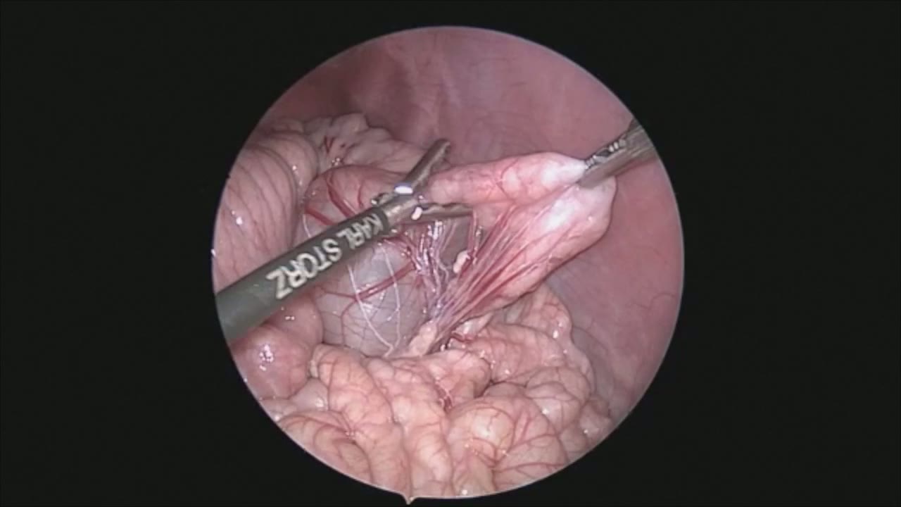
Laparoscopic Mesenteric and Short Gastric Dissection with a Vessel Sealer
Dr. Steve Rothenberg · 3 min · Published May 2026
Video
Laparoscopic Pyloromyotomy with a Reusable Handle and Disposable Blade
Dr. Steve Rothenberg · Published May 2026
Video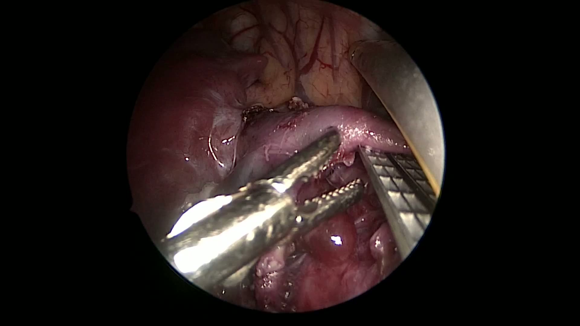
Thoracoscopic Stapling of the Left Lower Lobe Pulmonary Artery with a 5 mm Stapler
Dr. Steve Rothenberg · 9 s · Published May 2026
Video
Laparoscopic Plication of a Right Diaphragmatic Eventration in a 3-Month-Old
Dr. Steve Rothenberg · 6 min · Published May 2026
Video
Laparoscopic Redo Nissen Fundoplication in a 30 kg Child with the 3 mm Percuvance System
Dr. Steve Rothenberg · 5 min · Published May 2026
Video
Open Bowel Anastomosis
Dr. Steve Rothenberg · Published May 2026
Video
Intestinal Atresia Types Explained: Grossfeld Classification for Pediatric Surgery
1 min · Published Sep 2026
Video
Derivation and validation of the Pediatric Community-Acquired Pneumonia Severity (PedCAPS) score: A prospective cohort study
50 s · Published Sep 2026
Video
FETO for Late-Diagnosed Severe Congenital Diaphragmatic Hernia (CDH) at Cincinnati Children's with Dr. Beth Rymeski
4 min · Published Sep 2026
Video
Severe Congenital Diaphragmatic Hernia (CDH) Case: FETO Management & Unexpected Findings with Dr. Beth Rymeski
3 min · Published Sep 2026
Video
Fetoscopic Endoluminal Tracheal Occlusion (FETO) Procedure: Step-by-Step Guide with Dr. Beth Rymeski
4 min · Published Sep 2026
Video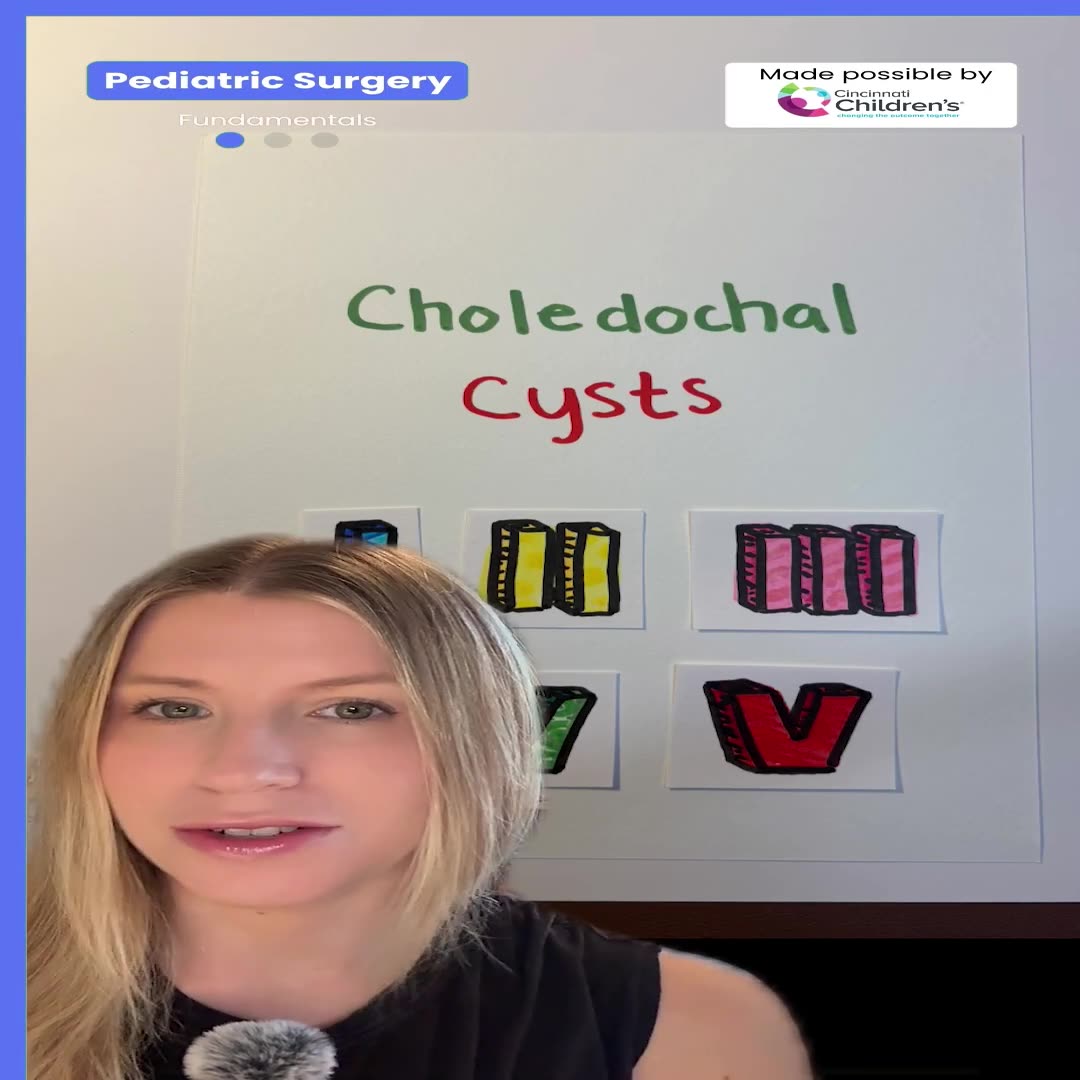
Choledochal Cyst Types Explained: Pediatric Surgery Fundamentals
1 min · Published Sep 2026
What the experts said
The first thoracoscopic TEF Type C repair was performed in 2000.
Single-lung ventilation is not required for thoracoscopic TEF repair; CO₂ insufflation at low flow (1 L) and low pressure (4–6 mmHg) is sufficient.
End-tidal CO₂ increases during thoracoscopic TEF repair, and anesthesiologists must accept CO₂ levels in the high 40s to low 50s mmHg.
Anesthesiologists should increase ventilation rate rather than tidal volume or peak inspiratory pressure to manage elevated CO₂, as increased pressures will re-expand the lung and obstruct the surgical field.
All babies desaturate initially when the lung is collapsed, but most recover within a couple of minutes.
Babies with congenital heart disease require specific cardiac anesthesia for thoracoscopic TEF repair.
Modified patient positioning at 45° prone (rather than full lateral) allows gravity to retract the lung, which is the biggest obstacle in thoracoscopic TEF repair.
99% of thoracoscopic TEF repairs can be performed using only ports without additional incisions.
The scope should be positioned far enough posterior to look down into the posterior mediastinum rather than up over the horizon, reproducing the view of an open thoracotomy.
The smallest patient operated on thoracoscopically for TEF repair was 1 kg, requiring modified port placement.
The scope port should be positioned just below and behind the tip of the scapula, with the left hand port just below and slightly behind the scope port so that right and left hands are at 90° during the critical anastomosis portion.
A 30° scope is used for thoracoscopic TEF repair.
The front row of anastomotic sutures is placed extraluminally.
The azygos vein is divided using vessel sealing devices to prevent bleeding during suturing, though some surgeons preserve it believing it may reduce stricture rates.
Vessel sealing devices are safer than monopolar cautery for dissection because there is no energy spread and less risk of surrounding tissue injury.
The vagus nerve should be preserved during fistula dissection.
In very small patients where a 5 mm clip cannot be used, the fistula is suture-ligated instead.
Clip ligation of the fistula is the fastest method to restore ventilation, as anesthesiologists have difficulty ventilating patients until the fistula is controlled due to significant tidal volume loss through large fistulas.
Trifurcation fistulas (entering at the carina) tend to create longer gaps than typical Type C fistulas.
Type C esophageal atresias are not considered long-gap defects.
The plane between the esophageal pouch and membranous trachea is easier to visualize thoracoscopically than through open thoracotomy.
When the upper pouch is densely adherent to the trachea, dissecting higher on the esophageal pouch makes the plane between esophagus and trachea more apparent.
Upper pouch fistulas, when present, are identified during bronchoscopy performed before pure esophageal atresia repairs.
The membranous tracheal wall cannot be visualized as well through open thoracotomy even with loupes as it can be seen thoracoscopically.
In thoracoscopic TEF repair, the most difficult part is avoiding injury to the membranous trachea, as there is sometimes almost a common wall between the pouch and trachea.
The tip of the upper pouch is where it is most adherent to the trachea, with almost no real plane present; the plane becomes easier to identify higher up on the pouch.
At the point where the upper pouch and trachea share a common wall, the safest dissection plane is submucosal on the esophagus to avoid entering the trachea.
Pediatric surgeons have an incredibly high rate of recurrent laryngeal nerve injuries.
The left recurrent laryngeal nerve is located just on the other side of the esophagus-trachea junction and should be identified and pushed away; it is closer to the esophagus than the trachea, especially higher up.
The magnified thoracoscopic view allows easier identification of the recurrent laryngeal nerve compared to open surgery.
All H-type fistulas are now repaired thoracoscopically because the nerve can be better identified than through a neck incision with blunt dissection.
With blunt dissection, there is minimal danger of damaging the recurrent laryngeal nerve.
The fistula is not divided immediately but left in place until ready for ligation, because dividing it early leads to difficulty finding it later.
In longer-gap repairs, the distal fistula is mobilized all the way down to the diaphragm without breaching the hiatus.
The tip of the upper pouch is completely amputated rather than just slit to create a good full-thickness opening, which may reduce stricture rates.
A bucket handle of tissue may be left on the upper pouch to provide a grasping point during suturing without injuring the tracheal wall.
The anastomosis is performed with a single layer of 5-0 PDS sutures on a TF needle, with the back wall sewn first starting with a middle stitch.
Large full-thickness bites including all layers are taken during anastomosis.
The entire anastomosis takes place in approximately a 2 square centimeter area.
When the gap is minimal, all knots are tied intracorporeally; for longer gaps, extracorporeal ties with a knot pusher allow watching the ends come together to ensure no tearing.
The back row of anastomotic sutures is placed intraluminally, typically consisting of 3 or 4 sutures.
Approximately 80% of surgeons can acquire the skill set to perform thoracoscopic TEF repair.
All surgeons in the speaker's practice perform thoracoscopic TEF repair after going through a training process.
Almost all surgeons can perform thoracoscopic TEF repair, though perhaps not to the highest technical level; the challenge is implementing training in an institution for a procedure that is not electively scheduled and does not occur frequently.
Training surgeons to perform thoracoscopic TEF repair requires commitment from experienced surgeons to operate with those learning, with very few contraindications to the approach except possibly patients on oscillators or with severe cardiac issues.
Three junior faculty members were trained to perform thoracoscopic TEF repair over approximately 3 years.
An NG tube is placed down about one-third of the way into the anastomosis as an internal stent to allow placement of the front row of sutures without closing off the lumen.
Five knots are thrown when using PDS suture for the anastomosis.
5-0 Vicryl can be used instead of PDS for the anastomosis and may be easier to teach.
PDS is preferred over Vicryl because it does not become wet and limp, maintains a more rigid tip for easier grasping in small spaces, and is a monofilament that does not soften as much as braided Vicryl.
NG tubes are no longer routinely left postoperatively based on discussion in the Midwest Consortium study, unless there is concern about anastomotic integrity requiring a stent.
Contrast studies are still performed on day 4 before starting feeds in the speaker's practice.
At 6-7 months postoperatively, thoracoscopic TEF repair results in three small 3-4 mm incisions with no chest wall deformity or musculoskeletal problems.
In the speaker's personal series, the leak rate is 3.5-4%, with no leaks requiring reoperation.
There has been one recurrent fistula in the speaker's series.
The stricture rate for Type C repairs is 14%, defined as strictures requiring intervention such as dilation.
The stricture rate in long-gap repairs is approximately 50%.
About 25% of patients undergoing thoracoscopic TEF repair eventually require fundoplication, with the rate being approximately 50% in pure esophageal atresia cases.
Only two aortopexies have been required in the speaker's series of thoracoscopic TEF repairs.
Chest drains are no longer routinely placed after thoracoscopic TEF repair if the repair is good and there are no concerns, based on experience from Great Ormond Street Hospital where contrast studies are not obtained, patients are fed on day 2, and chest drains are never left.