


From
StayCurrentMD
Technique: Laparoscopic Assisted Pull-through for Hirschsprung's Disease
With Dr. Steve Rothenberg
Chapter 2 of 6 · Surgical Management
Mesenteric dissection
Laparoscopic mesenteric dissection to pelvic floor
Cued at 1:21 · press play
Expert statements on this page
No expert statements were drawn from this page.
Host summaries · secondary, not cited in answers
Educational content from recorded physician discussions — not medical advice. Talk to your (or your child's) care team about your situation.
Video
Hirschsprung-Associated Enterocolitis: Recognition and Treatment
Nelson G. Rosen · 2 min · Published Jul 2019
Video
Rectal Atresia - a Unique Anorectal Malformation
4 min · Published Oct 2025
Video
How is Hirschsprung's Disease diagnosed? An ERNICA animation for parents and families
3 min · Published Dec 2023
Video
Quick Literature Updates Ep 23
4 min · Published Oct 2025
Video
Transition of Care - A general overview by Silja Kosolove
21 min · Published Nov 2023
Video
Transitional Care in Anorectal Malformation and Hirschsprung's Disease
1 min · Published Jul 2024
Video
Validation of an anorectal malformation trainer - Can a high-fidelity model simulate real life?
1 min · Published Jun 2026
Video
Complications and Long-Term Outcomes of Patients With Cloacal Malformation After Bowel Neovagina...
1 min · Published Jun 2026
Video
Safety and utility of long-acting steroid injection for management of post-operative stricture...
1 min · Published Jun 2026
Video
Association Between Social Determinants of Health and Choice of Urinary Reconstruction in Children
1 min · Published Jun 2026
Video
The Perineal Body Preserving PSARP (PPP)
11 min · Published Jun 2026
Video
Update Course Rewind 2025: Hirschsprung + ARM: Rare but Real
1 min · Published May 2026
Video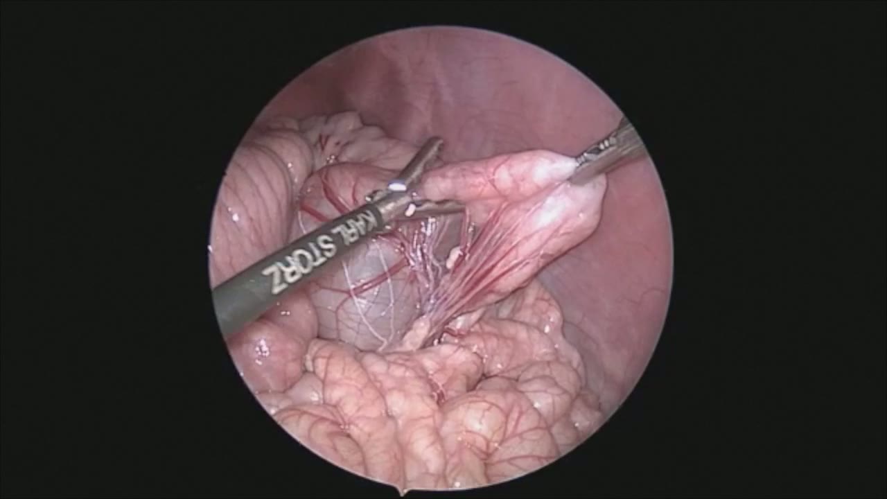
Laparoscopic Mesenteric and Short Gastric Dissection with a Vessel Sealer
Dr. Steve Rothenberg · 3 min · Published May 2026
Video
Laparoscopic Pyloromyotomy with a Reusable Handle and Disposable Blade
Dr. Steve Rothenberg · Published May 2026
Video
Thoracoscopic Stapling of the Left Lower Lobe Pulmonary Artery with a 5 mm Stapler
Dr. Steve Rothenberg · 9 s · Published May 2026
Video
Laparoscopic Plication of a Right Diaphragmatic Eventration in a 3-Month-Old
Dr. Steve Rothenberg · 6 min · Published May 2026
Video
Laparoscopic Redo Nissen Fundoplication in a 30 kg Child with the 3 mm Percuvance System
Dr. Steve Rothenberg · 5 min · Published May 2026
Video
Open Bowel Anastomosis
Dr. Steve Rothenberg · Published May 2026
Podcast
Diaphragm pacing as alternative to mechanical ventilation in pediatric respiratory failure
1 min · Published Nov 2026
Podcast
Severe CDH case with FETO: unexpected anatomic findings and clinical outcome
3 min · Published Nov 2026
Podcast
FETO procedure for late-diagnosed severe CDH with liver herniation
4 min · Published Nov 2026
Podcast
Pediatric pancreatitis: diagnosis, causes, and genetic insights with Dr. Juan Gurria
3 min · Published Nov 2026
Podcast
Sacral nerve stimulation for refractory pediatric fecal incontinence
2 min · Published Nov 2026
Podcast
Fetoscopic endoluminal tracheal occlusion for congenital diaphragmatic hernia
4 min · Published Nov 2026
What the experts said
The key to mesenteric dissection is staying exactly adjacent to the colon wall to prevent injury to surrounding structures.
Because all heat and energy remains between the jaws of the sealer, there is no danger in injuring surrounding structures by brushing aside them with the sealer.
Small perforating vessels are grasped, sealed, and then pulled down off the rectal wall during circumferential dissection.
Dissection at the peritoneal reflection uses the same technique of grasping the peritoneal reflection and small perforating vessels and pulling them down off the colon.
The 3 millimeter sealer allows dissection of vessels and mobilization of tissue around the colon without risk of pass-pointing, unlike the 3 millimeter hook which was previously the preferred mode of dissection.
Using the 3 mm sealer eliminates the need for instrument changes with the right hand throughout the case.
Carrying the laparoscopic dissection down to the pelvic floor limits the amount of transanal dissection necessary and decreases the risk of injury to the external sphincter.
Because there is no energy spread from the tips of the 3 mm sealer, it is safe for the instrument to be adjacent to the bladder, vagina, prostate, and other surrounding structures.
Energy being only between the jaws of the instrument diminishes the risk of injury to the ureters, vas deferens, and other vital structures.
The preferred timing is to perform this operation in the newborn period prior to discharge to home, though it is acceptable if the child tolerates rectal irrigations to let them grow.
With current technology, the operation is considered extremely safe in the newborn period.
For the transanal dissection, traction stitches are placed inside the anus just proximal to the dentate line and then out to the skin, slightly inverting the anus so the dentate line can be clearly visualized.
Between 4 to 8 sutures are used to evert the anus.
A mucosal incision is made with hand cautery 2 to 3 millimeters proximal to the dentate line and crypt.
The key to the transanal portion is that it should all take place externally to the anus.
The laparoscopic dissection down to the pelvic floor allows the transanal dissection to be carried out outside of the anus, so no retractors are ever placed within the external sphincter which may cause these muscles to be damaged.
Sharp dissection is preferred to mobilize the submucosal space, although blunt dissection can also be used.
Performing the dissection external to the anus protects the external sphincter muscles and improves the chance of good continence.
The colon is divided 5 to 6 centimeters above the biopsy site, which is 5 centimeters above the obvious transition zone.
The coloanal anastomosis is performed with 12 to 16 sutures total, placing 3 to 4 additional sutures in each quadrant after the initial 4 quadrant stay sutures.
Vicryl suture is used for the anastomosis in newborns.
This operation took 70 minutes.
The child was left without a nasogastric tube and started stooling the morning following surgery.
The patient was started on feeds less than 24 hours after the procedure.
The anastomosis is calibrated with a 12 Hegar dilator at the end of the procedure.
Gauze packing is placed in the anus at the end of the procedure.
The JRS 3 millimeter sealer is used as the primary mode of dissection for laparoscopic-assisted pull-through in Hirschsprung's disease.
The patient was a newborn weighing 3.2 kg.
A super umbilical ring incision is used for small newborns to place the Veress needle and 4 mm trocar, with care taken to avoid the umbilical vein by placing the trocar just to the left of midline.
A full thickness biopsy is obtained from the muscular wall approximately 5 centimeters above the transition zone between the distal sigmoid and rectum.