
Staged Closure of Gastroschisis with Spring-loaded Silo
Educational content from recorded physician discussions — not medical advice. Talk to your (or your child's) care team about your situation.
Video
Staged Closure of Gastroschisis with Spring-loaded Silo
27 min · Published Feb 2020
Video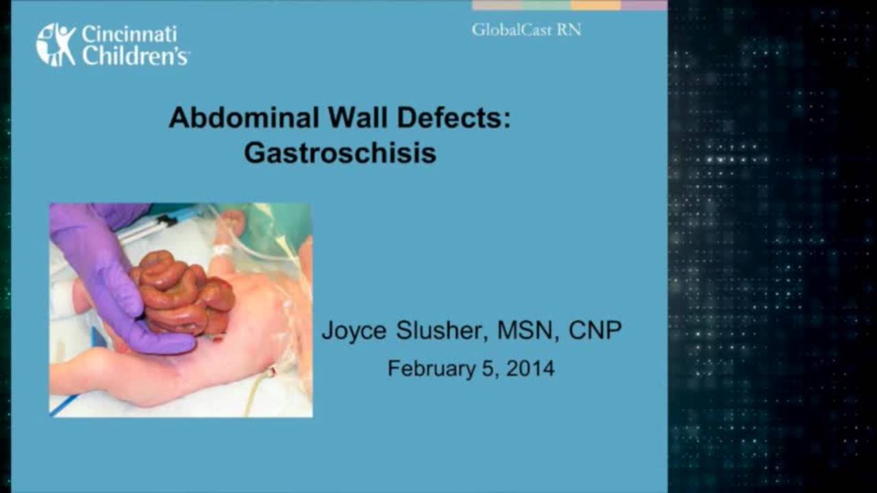
Gastroschisis: Advanced Practice Providers
43 min · Published Feb 2014
Video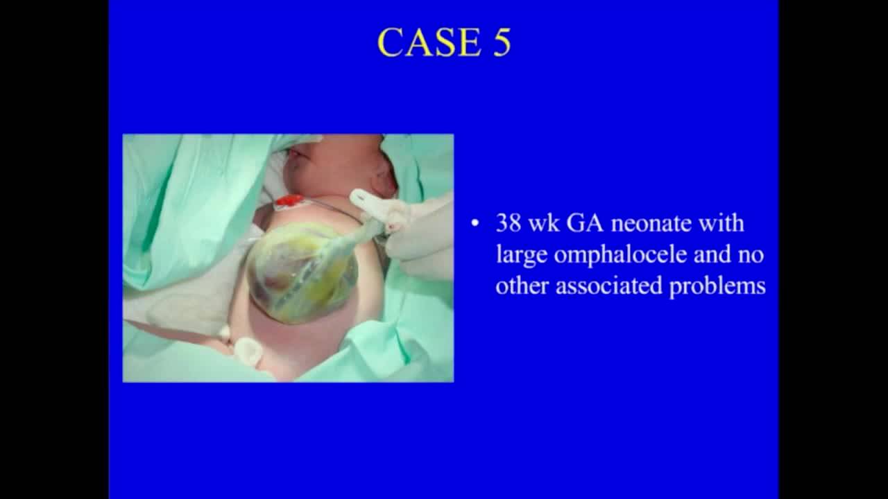
Abdominal Wall Defects
37 min · Published Nov 2018
Video
Abdominal Wall Defects: Update Course 2013
37 min · Published Sep 2013
Video
Omphalocele & Gastroschisis
Dr. Todd Ponsky · 9 min · Published Mar 2022
Podcast
Omphalocele and Gastroschisis With Dr. Foong-Yen Lim
9 min · Published Feb 2022
Video
Pediatric Surgical Oncology Research Collaborative (PSORC): Studying Rare Pediatric Tumors
56 s · Published May 2026
Video
Update Course Rewind 2025: Hirschsprung + ARM: Rare but Real
1 min · Published May 2026
Video
Update Course Rewind 2025: Hirschsprung + ARM: Rare but Real
1 min · Published May 2026
Video
Pooling Patients to Study Rare Pediatric Tumors: An Introduction to PSORC
56 s · Published May 2026
Video
The fetal frontier: A review of current and emerging fetal therapies for genetic diseases
44 s · Published May 2026
Video
Indocyanine green assists with sentinel lymph node mapping in pediatric and adolescent patients
1 min · Published May 2026
What the experts said
At UCI pediatric surgery practice serving 2 neonatal intensive care units, 30 to 40 patients per year with gastroschisis are treated, making it second only to inguinal hernias as a congenital anomaly requiring surgical correction.
For the last 5 years on the UCI pediatric surgery service, spring-loaded silos have been routinely placed for patients with gastroschisis with excellent results.
Most babies with gastroschisis are delivered vaginally after spontaneous onset of labor; routine cesarean section is not performed, nor is early labor induced.
For silo placement, the baby is sedated with fentanyl and midazolam drips, intubated, and given a single dose of vecuronium; while placement is feasible without these interventions, they create optimal conditions and a well-controlled situation.
A ring that is 2 centimeters larger than the diameter of the defect is typically chosen; since most defects are 2 to 3 centimeters in diameter, 4 and 5 centimeter silos are most commonly used.
Gastroschisis cases involving an atresia typically contain severely distended bowel and often require a 7.5 centimeter silo.
Evacuation of the colon is important as it will significantly decrease the size of the colon and allow for faster reduction.
If an obstruction exists without perforation, the policy is to proceed with silo placement and closure, followed by exploration 4 to 6 weeks later.
Final closure is performed when the silo contents is within 2 centimeters of the abdominal wall.
The silo is left in place for the shortest time possible; unnecessary prolongation has no advantages, may make closure more involved by slowly enlarging the defect, and may increase infectious and other potential complications.
Stretching the abdominal wall aids in a tension-free closure and often produces some minor postoperative congestion of the abdominal wall.
For skin closure, bites are taken approximately 3 millimeters from the skin edge because the edge is often slightly ischemic and bites exactly in the skin edge are likely to cause skin necrosis and possible wound infection.
The Bentek silo can be used for staged reduction of omphalocele after excision of the sac, with the liver gradually reduced along with the bowel.
When a large silo is required for a prolonged period, a few corner stitches between the silo ring and the abdominal wall prevent premature dislodgement of the silo.
The silo can be used in cases of neonatal abdominal compartment syndrome, such as in severe diffuse necrotizing enterocolitis.
At the University of California Irvine, this method is applied to all patients with gastroschisis and selectively used in other situations where abdominal wall closure is not possible.
Gastroschisis has a reported incidence of 1 in 6000 to 1 in 10,000, but is much more common in many parts of the US including Southern California.
Traditional staged closure with sutured elastic silo involves risks of silo disruption, fascial dehiscence, and infectious complications.
The spring-loaded silo allows for fast, pain-free, sutureless silo placement without need for a formal operation.
Doctor James Fisher and colleagues from Loma Linda University were the first to publish a series of patients to undergo routine silo placement at the bedside.
Several retrospective studies from large US centers reported that patients who underwent routine silo placement with delayed closure showed one or more advantages: decreased airway pressures, earlier extubation, decreased incidence of necrotizing enterocolitis, decreased infectious complications, more rapid return of bowel function, decreased length of stay, and decreased hospital charges.
The Bentek silo is available in 7 sizes from 3 centimeters to 15 centimeters, defined by the diameter of the reinforced ring.
A distal colon severely distended with meconium is typical and is a good indication of the probable absence of a proximal atresia or stenosis.
If the ring has too much traction it will cause abdominal wall congestion and edema, complicating closure later on.
Too little traction will allow the ring to exert constant pressure on the abdominal contents, most notably the duodenum, with a risk of pressure necrosis.
The silo creates a closed system by completely containing the bowel and peritoneal fluid.
A 1 centimeter fascial edge is mobilized in both directions to the border of the umbilical stump.
The baby remained stable without any increase in airway pressures during the closure procedure, which was completed in under 25 minutes.
Essential factors for optimal outcome include: appropriate choice of silo size, avoiding mesenteric torsion, final closure when near complete reduction is achieved and not prolonging silo duration, close observation of silo configuration and contents, purse string closure of fascia and skin, and preservation of the umbilical stump.