27 views 0 likes

StayCurrentMD

GCMD Space · View profile →

Staged Closure of Gastroschisis with Spring-loaded Silo

Video Published 2020-02-24 Updated 2026-08-01

Timestops (8)

Topic Overview

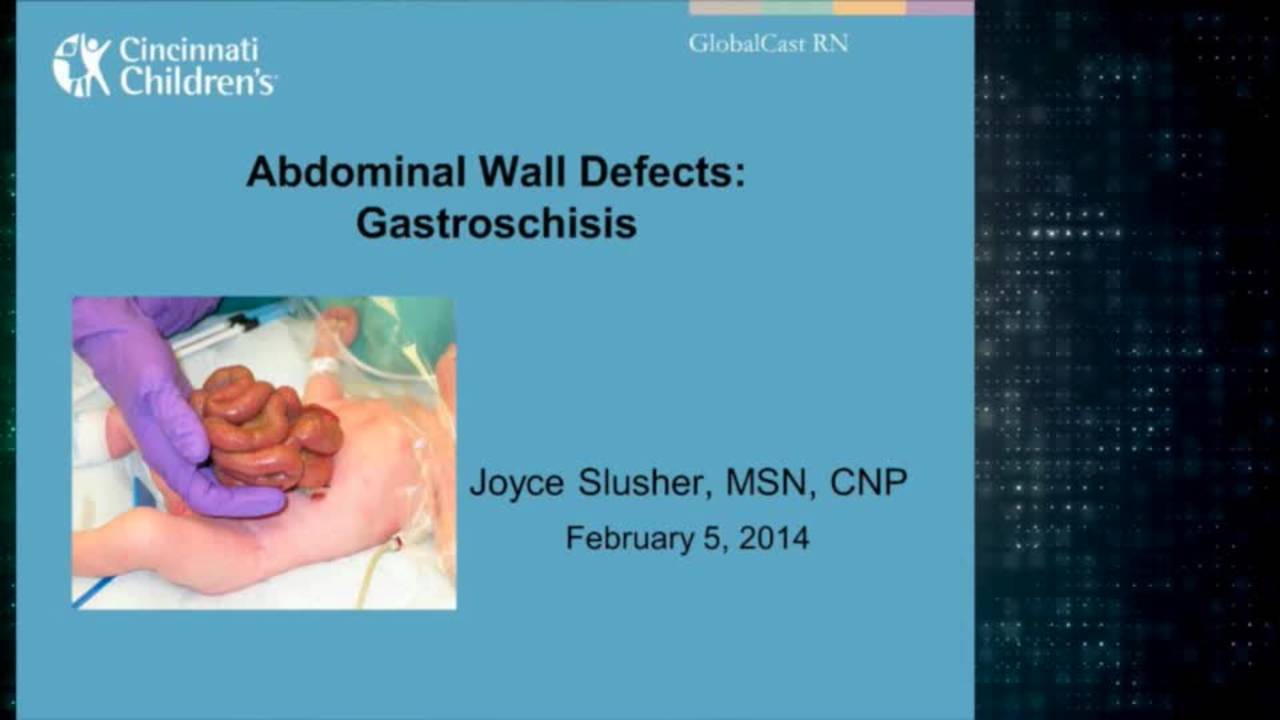
This educational video demonstrates the staged closure technique for gastroschisis using a spring-loaded silo at UC Irvine Medical Center. The presentation covers the epidemiology of gastroschisis (reported incidence 1 in 6,000-10,000, though more common in Southern California), the evolution from traditional primary closure to routine staged closure, and detailed technical aspects of silo placement and subsequent fascial closure. Multiple retrospective studies have shown advantages of staged closure including decreased airway pressures, earlier extubation, decreased necrotizing enterocolitis, and shorter hospital stays, though prospective randomized data are pending. The technique involves bedside silo placement without sutures, gradual reduction over several days, and delayed fascial closure using a purse-string technique.

Key Takeaways

  • Spring-loaded silos enable sutureless bedside placement without formal operation, avoiding risks of traditional sutured silo dehiscence.
  • Routine staged closure with spring-loaded silos shows decreased NEC, faster extubation, earlier bowel function return vs. primary closure.
  • Choose silo ring 2cm larger than defect diameter; decompress distended colon/stomach/bladder before placement to optimize reduction.
  • Most gastroschisis defects are 2-3cm, requiring 4-5cm silos; complex cases with atresia/severe peel may need 7.5cm silos.
  • Sedation, intubation, and single-dose paralytic create optimal controlled conditions for silo deployment despite feasibility without.

Inside this episode

Kai, the Library's AI content creator, listened to this episode and mapped who's speaking, the chapters, key claims, and cases. Every item links to the exact moment in the recording.

AI-enriched

Who's speaking

  • Speaker 1 — host
  • Speaker 2
  • Speaker 3

Chapters

  • 0:26Introduction and Background on Gastroschisis Management — Overview of gastroschisis epidemiology, traditional vs. staged closure approaches, introduction of spring-loaded silo technology, and published outcomes data.
  • 3:46Silo Selection and Pre-Procedure Preparation — Objectives outlined, prenatal management described, delivery room care, NICU preparation with sedation and intubation, and principles of choosing appropriate silo size.
  • 6:38Silo Placement Technique — Step-by-step demonstration of bedside silo placement including bowel examination, meconium evacuation, silo preparation, bowel reduction into silo, and ring deployment under fascia.
  • 11:52Silo Management and Reduction Process — Post-placement care including suspension technique, daily reduction maneuvers, monitoring for complications, and criteria for final closure.
  • 14:00Final Fascial Closure Technique — Operating room procedure for definitive closure including silo removal, fascial mobilization, purse-string fascial closure, and skin closure with neo-umbilicus creation.
  • 23:48Outcomes, Key Principles, and Alternative Applications — Cosmetic results at follow-up, summary of essential technical factors, and examples of silo use in other conditions including bowel gangrene, omphalocele, abdominal wall agenesis, and necrotizing enterocolitis.

Key claims

  • 0:32Gastroschisis is a full thickness abdominal wall defect that typically occurs to the right of the umbilicus — Speaker 1
  • 0:39Gastroschisis has a reported incidence of 1 in 6000 to 1 in 10,000 — Speaker 1
  • 0:49At UC Irvine's two neonatal intensive care units, they care for 30 to 40 patients per year with gastroschisis — Speaker 1
  • 0:57Gastroschisis is second only to inguinal hernias as a congenital anomaly requiring surgical correction at their institution — Speaker 1
  • 1:11Traditional staged closure with sutured elastic silo carries risks of silo disruption, fascial dehiscence, and infectious complications — Speaker 1
  • 1:27The spring-loaded silo allows for fast, pain-free, sutureless silo placement without need for a formal operation — Speaker 1
  • 2:08Dr. James Fisher and colleagues from Loma Linda University were the first to publish a series of patients undergoing routine bedside silo placement — Speaker 1
  • 2:33A prospective randomized study comparing primary to staged closure of gastroschisis is in progress but results are not yet available — Speaker 1
  • 2:43Several retrospective studies from large US centers reported favorable results with spring-loaded silo staged closure compared to primary closure controls — Speaker 1
  • 2:57Advantages of routine silo placement with delayed closure include decreased airway pressures, earlier extubation, decreased incidence of necrotizing enterocolitis, decreased infectious complications, more rapid return of bowel function, decreased length of stay, and decreased hospital charges — Speaker 1
  • 4:30Most babies with gastroschisis are delivered vaginally after spontaneous onset of labor; routine cesarean section is not performed nor is early labor induced — Speaker 1
  • 5:20The Bentek silo is available in 7 sizes from 3 centimeters to 15 centimeters diameter — Speaker 1
  • 5:37A ring that is 2 centimeters larger than the diameter of the defect is typically chosen — Speaker 1
  • 5:42Most gastroschisis defects are 2 to 3 centimeters in diameter, making 4 and 5 centimeter silos the most commonly used — Speaker 1
  • 5:59Gastroschisis cases involving atresia typically contain severely distended bowel and often require a 7.5 centimeter silo — Speaker 1
  • 6:56A distal colon severely distended with meconium is a good indication of the probable absence of a proximal atresia or stenosis — Speaker 1
  • 8:09If an obstruction exists without perforation, their policy is to proceed with silo placement and closure, followed by exploration 4 to 6 weeks later — Speaker 1
  • 13:02Too much traction on the ring will cause abdominal wall congestion and edema, complicating closure later — Speaker 1
  • 13:09Too little traction will allow the ring to exert constant pressure on abdominal contents, most notably the duodenum, with risk of pressure necrosis — Speaker 1
  • 13:20Final closure is performed when the silo contents are within 2 centimeters of the abdominal wall — Speaker 1
  • 13:34Unnecessary prolongation of silo duration has no advantages, may make closure more involved by slowly enlarging the defect, and may increase infectious and other potential complications — Speaker 1
  • 13:52The silo essentially creates a closed system by completely containing the bowel and peritoneal fluid — Speaker 1
  • 21:07Skin edge bites exactly at the edge are likely to cause skin necrosis and possible wound infection; bites should be approximately 3 millimeters from the skin edge — Speaker 1
  • 25:31The Bentek silo can be used for staged reduction of omphalocele after excision of the sac, with gradual reduction of liver along with bowel — Speaker 1
  • 26:10When a large silo is required for a prolonged period, a few corner stitches between the silo ring and abdominal wall prevent premature dislodgement — Speaker 1
  • 26:34The silo can be used in cases of neonatal abdominal compartment syndrome such as severe diffuse necrotizing enterocolitis — Speaker 1

Cases discussed

  • 6:14Gastroschisis with mild evisceration and minimal distention
  • 6:14Gastroschisis with severe evisceration
  • 12:48Gastroschisis with near complete reduction in 6 days
  • 13:48Gastroschisis reduced in 4 days
  • 14:056-day-old infant undergoing final fascial closure
  • 25:09Gastroschisis with bowel segment of tenuous blood supply
  • 25:51Abdominal wall agenesis with massive herniation
  • 26:34Neonatal abdominal compartment syndrome from necrotizing enterocolitis

Open questions

  • What will be the results of the prospective randomized study comparing primary to staged closure of gastroschisis?
This episode was analyzed and enriched by Kai, the Library's AI content creator. Every item links to the moment it comes from — click a timestamp to listen in context.
Written for:

Spring-Loaded Silo for Gastroschisis: Staged Closure Without Sutures

The episode's main topic retold as a plain-language walkthrough — what it is, why it matters, and what the speakers concluded. Written by Kai from the episode transcript and reviewed before publishing.

For the care team · Explainer · AI-written, human-reviewed

Why This Technique Exists

Gastroschisis — a full-thickness abdominal wall defect typically to the right of the umbilicus 0:32 — is common enough that a busy pediatric surgery service may see thirty to forty cases per year 0:49. For decades, surgeons attempted immediate primary closure in the delivery room or operating room, resorting to staged closure only when the bowel could not be safely reduced. Traditional staged closure required suturing an elastic silo to the fascial edges, carrying risks of silo disruption, fascial dehiscence, and infection 1:11. The spring-loaded silo eliminated the need for sutures, anesthesia, and a formal operation, allowing bedside placement in the NICU 1:27. This shift — pioneered by Fisher and colleagues at Loma Linda 2:08 — has made staged closure the default approach at many centers.

The Core Problem

The eviscerated bowel in gastroschisis is edematous, often coated in inflammatory peel, and may be distended with meconium. Forcing it back into a small abdominal cavity immediately after birth raises intra-abdominal pressure, compromises ventilation, and risks bowel ischemia. The alternative — gradual reduction over several days — allows the abdominal domain to accommodate the viscera without physiologic compromise. Retrospective series report advantages including decreased airway pressures, earlier extubation, lower rates of necrotizing enterocolitis, faster return of bowel function, and shorter hospital stays 2:57. A prospective randomized trial comparing primary to staged closure is underway but results are not yet available 2:33.

How the Approach Works

The spring-loaded silo is a transparent silastic cylinder attached to a compressible metal-reinforced ring 1:27. The ring is available in seven sizes from 3 to 15 centimeters diameter 5:20. Most gastroschisis defects measure 2 to 3 centimeters, so 4- and 5-centimeter silos are most commonly used 5:42; a ring 2 centimeters larger than the defect diameter is typical 5:37. Cases involving atresia, where the bowel is severely distended, often require a 7.5-centimeter silo 5:59.

Most infants are delivered vaginally after spontaneous labor — routine cesarean section or early induction is not performed 4:30. After delivery, the eviscerated contents are wrapped in moist gauze or a bowel bag and the infant is transported to the NICU. The infant is sedated, intubated, and given a single dose of neuromuscular blockade to create optimal conditions for silo placement 1:27. The bowel is examined for atresia, stenosis, ischemia, or perforation. A distal colon severely distended with meconium is a favorable sign, indicating probable absence of proximal obstruction 6:56. The colon is evacuated to reduce its bulk. If an obstruction exists without perforation, the policy is to proceed with silo placement and closure, then explore four to six weeks later rather than operating immediately 8:09.

The silo ring is compressed, inserted into the defect, and released. It immediately regains its spherical shape and stays in position without sutures. The bowel is reduced into the silo, and the ring is palpated to confirm no loops are trapped beneath it. The silo is suspended from the bed so that the ring remains just palpable under the fascia but does not elevate the abdominal wall. Too much traction causes abdominal wall congestion and edema, complicating later closure 13:02. Too little traction allows the ring to exert constant pressure on underlying structures — most notably the duodenum — with risk of pressure necrosis 13:09. The silo essentially creates a closed system, completely containing the bowel and peritoneal fluid 13:52.

Daily reduction is performed by gently compressing the silo and advancing the bowel into the abdomen. Final closure is performed when the silo contents are within 2 centimeters of the abdominal wall 13:20. Unnecessary prolongation has no advantages, may slowly enlarge the defect, and increases the risk of infectious complications 13:34.

The Closure Procedure

Definitive closure is performed in the operating room. The silo is removed and the fascia is mobilized circumferentially by raising a skin and subcutaneous flap, starting opposite the umbilicus. A running purse-string suture of PDS is placed around the fascial edge with alternating inside-out and outside-in bites. The suture is tightened gradually while an assistant compresses the abdominal wall to relieve tension, creating a tension-free closure. The skin is closed with a running monocryl suture, taking bites approximately 3 millimeters from the edge — not exactly at the edge, which is often ischemic and prone to necrosis 21:07. The running stitch purse-strings the skin to create a neo-umbilicus.

Beyond Gastroschisis

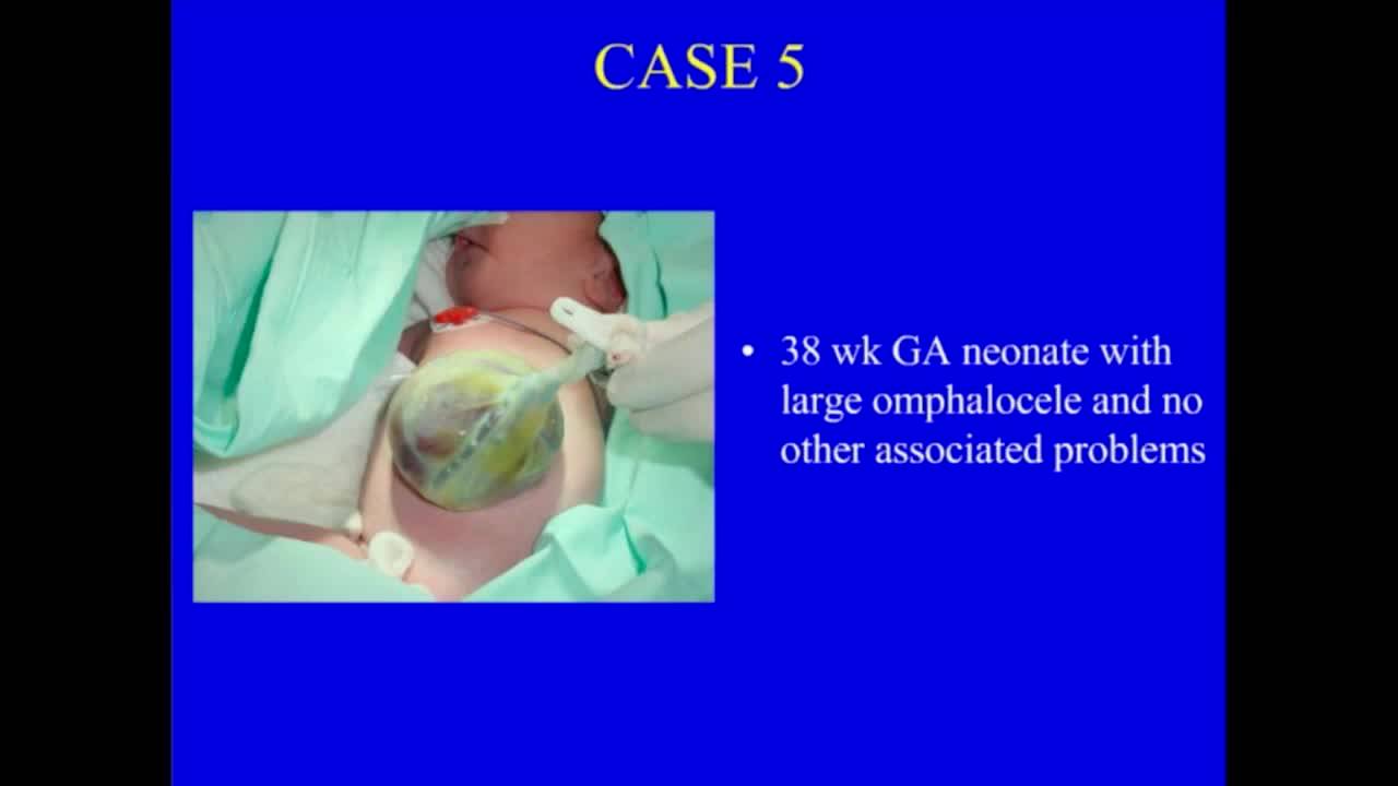
The same silo can be used for staged reduction of omphalocele after excision of the sac, with gradual reduction of liver along with bowel 25:31. In cases requiring a large silo for a prolonged period, a few corner stitches between the ring and abdominal wall prevent premature dislodgement 26:10. The silo also has a role in neonatal abdominal compartment syndrome, such as severe diffuse necrotizing enterocolitis 26:34.

When to Involve Pediatric Surgery

Gastroschisis is typically diagnosed on prenatal ultrasound. Referral to a center with pediatric surgery and neonatal intensive care should occur as soon as the diagnosis is made. Delivery should be planned at that center to allow immediate postnatal management. The defect requires surgical closure within the first week of life.

Takeaways from this story

  • Spring-loaded silo allows bedside placement without sutures, anesthesia, or operating room, eliminating risks of traditional sutured silo.
  • Choose silo ring 2 cm larger than defect diameter; most defects are 2-3 cm, making 4-5 cm silos most common.
  • Silo suspension must keep ring just palpable under fascia — too much traction causes wall edema, too little risks duodenal pressure necrosis.
  • Close when silo contents are within 2 cm of abdominal wall; prolonging silo duration has no advantage and may enlarge defect or increase infection risk.
  • If obstruction exists without perforation, proceed with silo closure and explore 4-6 weeks later rather than operating immediately.

Keywords

Hashtags

Transcript

Comments

Loading comments…