16 views 0 likes

Dr. Todd Ponsky

Pediatric Surgery · View profile →

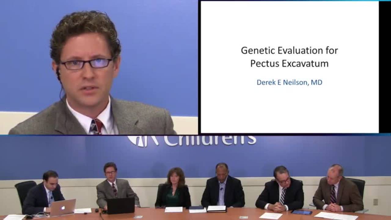
Pectus - Preoperative Assessment - Radiology and Cardiac Evaluation

Video Published 2018-11-10 Updated 2022-08-22

Timestops (6)

Topic Overview

A multidisciplinary discussion on preoperative assessment of pectus excavatum, emphasizing the shift from CT to cardiac MRI for evaluation. The panel presents data showing that approximately 15% of pectus patients have reduced right ventricular ejection fraction (below 50%), with compression of the RV free wall correlating with decreased function. The discussion addresses limitations of the Haller index in patients with barrel-shaped chests and introduces supplementary metrics (correction index, depression index) that may better identify surgical candidates. The team advocates for a holistic approach including genetics referral for connective tissue disorders and metal allergy testing, while debating optimal surgical timing and the functional versus cosmetic nature of the condition.

Key Takeaways

  • ~15% of pectus patients have RV ejection fraction <50%; compression of RV free wall correlates with worse function. (21:30)
  • Cardiac MRI replaces CT+echo: 25min scan, no contrast/radiation, detects RV compression echo misses, comparable cost. (16:42)
  • Haller index fails in barrel chests; correction index >10% and depression index >0.2 better identify surgical candidates. (13:57)
  • Bundling multiple metrics (Haller, correction, depression indices + cardiac dysfunction) effective for insurance approval. (31:08)
  • Screen all pectus patients for connective tissue disorders (genetics referral) and metal allergy; affects pain/outcomes. (6:32)

Inside this episode

Kai, the Library's AI content creator, listened to this episode and mapped who's speaking, the chapters, key claims, and cases. Every item links to the exact moment in the recording.

AI-enriched

Who's speaking

  • Todd — host
  • Dr. Vic Garcia — guest
  • Derek Nielsen — guest
  • Dr. Becky Brown — guest
  • Dr. Michael Taylor — guest
  • Dr. Eric Crotty — guest
  • Speaker 7

Chapters

  • 0:00Introduction and Faculty Presentation — Opening remarks, housekeeping, and introduction of the multidisciplinary faculty including surgery, cardiology, radiology, and genetics.
  • 4:26Case Presentation — 13-year-old male with pectus excavatum, Haller index 4.8, nickel allergy requiring titanium bars, developed chronic pain at 2 years post-op, highlighting the holistic assessment approach.
  • 7:34Radiology Assessment and MRI Protocol — Discussion of transition from CT to MRI, multiple measurement indices (Haller, correction, depression), and limitations of Haller index in barrel-chested patients.
  • 16:42Cardiac MRI Findings and RV Function — Presentation of cardiac MRI data showing 15% of patients have RV ejection fraction below 50%, correlation between RV compression and decreased function, advantages of MRI over echocardiography.
  • 29:30Clinical Implications and Insurance Considerations — Discussion of functional versus cosmetic debate, insurance requirements, bundling multiple metrics for approval, and the role of cardiac dysfunction in surgical decision-making.
  • 38:20Optimal Surgical Timing and Age Considerations — Debate on appropriate age for repair, audience poll results showing preference for 9-11 years, discussion of Dr. Park's experience with younger patients and asymmetric defects.

Key claims

  • 2:48Pectus excavatum repair is arguably the most painful operation that could be done in a child — Dr. Vic Garcia
  • 21:30Approximately 15% of pectus patients had right ventricular ejection fractions less than 50% (normal is 50%) — Dr. Michael Taylor
  • 23:25Patients with RV compression had depressed right ventricular ejection fractions — Dr. Michael Taylor
  • 23:30Patients with compression of the RV free wall versus the RA-RV groove had more decreased RV function — Dr. Michael Taylor
  • 24:31Kids can have RV ejection fractions in the 40s and be asymptomatic — Dr. Michael Taylor
  • 14:50The Haller index can be normal in patients with obvious pectus deformity due to barrel-shaped chest configuration — Dr. Eric Crotty
  • 13:57The correction index greater than 10% shows no overlap between normal and abnormal patients — Dr. Eric Crotty
  • 14:11Depression index greater than 0.2 is the cutoff between normal and abnormal patients — Dr. Eric Crotty
  • 12:18Haller's 3.25 cutoff was from an observational retrospective study, not a rigorous scientific prospective study — Dr. Eric Crotty
  • 16:42Cardiac MRI takes about 25 minutes and provides anatomical and functional evaluation without contrast — Dr. Michael Taylor
  • 18:34Echocardiogram image quality in most pectus patients is atrocious due to the deformity — Dr. Michael Taylor
  • 19:22MRI can detect the pectus compressing the anterior surface of the right ventricle, which is nearly impossible to see with echo — Dr. Michael Taylor
  • 19:47One cardiac MRI is comparable in cost to an echo plus CT in most places — Dr. Michael Taylor
  • 40:10Standard CT dose can deliver about 7 millisieverts of radiation, equivalent to 2 years of background radiation — Dr. Eric Crotty
  • 39:29Younger patients have higher long-term cancer risk from CT radiation due to more rapidly dividing cells — Dr. Eric Crotty
  • 41:13There is data suggesting increased risk of malignancy long term from even one CT scan — Dr. Eric Crotty
  • 31:08Insurance companies require evidence of cardiac dysfunction or abnormality in addition to pectus index for approval — Dr. Vic Garcia
  • 31:58Bundling multiple metrics (Haller, correction, depression indices plus cardiac dysfunction) has been effective in reversing insurance denials — Dr. Vic Garcia
  • 32:48When pectus orientation has sternum pressing on RV free wall, it affects right ventricular function — Dr. Michael Taylor
  • 28:15Children tolerate MRI very well, especially with video goggle technology to watch movies during the procedure — Dr. Eric Crotty
  • 29:09MRI can be performed with pectus bar in place with only minimal localized artifact — Dr. Michael Taylor
  • 22:20The pectus geometry is three-dimensional and varies dramatically in superior-inferior dimension — Dr. Michael Taylor
  • 26:20As kids age, the heart appears to get extruded into the left chest with pectus — Dr. Michael Taylor
  • 42:15Dr. Park in Korea argues that repair can be done safely in patients older than 3 years — Dr. Becky Brown
  • 42:31Bars that extend all the way to mid-axillary line can impede chest wall growth in younger patients — Dr. Vic Garcia
  • 43:01Overcorrection converting excavatum to carinatum can occur, particularly in patients with connective tissue disorders — Dr. Vic Garcia
  • 43:35Park reports seeing fewer asymmetric patients when correcting at younger ages — Dr. Vic Garcia
  • 6:32Patients with connective tissue disorders are at risk for chronic pain after pectus repair — Dr. Becky Brown
  • 6:43Metal allergy testing is performed in every pectus patient at this center — Dr. Becky Brown
  • 17:38Cardiac MRI can screen for valve disease and aortopathy associated with connective tissue disorders — Dr. Michael Taylor

Cases discussed

  • 5:3213-year-old male with pectus excavatum, tall thin habitus, hyperflexible, presenting with progressive exercise intolerance
  • 35:444-year-old with Marfan syndrome and Haller index of 36, presenting with progressive exercise intolerance

Points of disagreement

  • 25:14Functional versus cosmetic nature of pectus repair
    • Todd: Frequently sends patients away saying it's cosmetic, has pectus himself and is fine with it, questions whether measurements translate to actual clinical significance
    • Dr. Vic Garcia: Some colleagues at his institution feel it is cosmetic, but he believes the cardiac dysfunction data supports functional indication
  • 34:39Optimal age for pectus repair
    • 50% believe age must be greater than 9-11 years, 30% say over 5 years is acceptable
    • Dr. Vic Garcia: Dr. Park argues repair can be done safely in patients as young as 3 years old, with potential benefit of preventing asymmetric deformities
  • 30:37Necessity of imaging for pectus assessment
    • Questions need for CT or MRI when it doesn't change management, especially in patients with obvious pectus on exam
    • Dr. Vic Garcia: Imaging is necessary for insurance approval and to document cardiac dysfunction, provides bundle of evidence for reversing denials

Open questions

  • Does right ventricular ejection fraction in pectus patients change over time (e.g., from age 8 to 15) or does it remain stable/worsen?
  • At what age does cardiac extrusion into the left chest become irreversible, and is there a critical window for surgical correction?
  • What are the long-term cardiac outcomes in adults with uncorrected pectus excavatum and reduced RV function?
  • Can formal cardiopulmonary exercise testing better identify which asymptomatic pectus patients have true functional impairment?
  • Why does Dr. Park see fewer asymmetric pectus cases when operating at younger ages - does early correction prevent asymmetry development?
  • What is the optimal bar length and configuration for younger patients to avoid impeding chest wall growth while maintaining correction?
This episode was analyzed and enriched by Kai, the Library's AI content creator. Every item links to the moment it comes from — click a timestamp to listen in context.
Written for:

Preoperative Imaging for Pectus Excavatum: Why Cardiac MRI Replaced CT

The episode's main topic retold as a plain-language walkthrough — what it is, why it matters, and what the speakers concluded. Written by Kai from the episode transcript and reviewed before publishing.

For the care team · Explainer · AI-written, human-reviewed

Why This Exists

Pectus excavatum — the inward depression of the anterior chest wall — is the most common congenital chest wall deformity. For decades it was dismissed as cosmetic. That changed when surgeons recognized that the sternum can compress the right ventricle, and when minimally invasive repair (the Nuss procedure) made correction feasible in adolescence. But the operation is painful, the bars stay in for years, and insurance coverage hinges on proving physiologic impairment. A comprehensive preoperative assessment became necessary — not to justify surgery to skeptics, but to select the right patients and document the indication.

The Core Clinical Problem

The challenge is threefold. First, measure the severity of the deformity in a way that accounts for chest shape variability — barrel-chested patients can have severe pectus with a falsely normal Haller index 14:50. Second, determine whether the sternum is compressing the heart and whether that compression affects function. Third, do all of this without cumulative radiation exposure in children who may need serial imaging.

Traditionally this required CT for chest geometry and echocardiography for cardiac assessment. But echo image quality in pectus patients is poor — the sternum creates near-field artifact that obscures the right ventricle 18:34. And CT delivers approximately 7 millisieverts per study, equivalent to two years of background radiation, with evidence of increased long-term cancer risk even from a single scan 40:10 41:13. Younger patients face higher risk because their cells divide more rapidly 39:29.

How the Approach Works

Cardiac MRI replaced both studies. A single 25-minute scan provides chest wall geometry, cardiac anatomy, and ventricular function without contrast or radiation 16:42. The protocol measures three indices rather than relying on Haller alone. The Haller index (transverse diameter divided by anteroposterior diameter) was never rigorously validated — it came from a retrospective observational study, yet insurance companies adopted the 3.25 threshold as gospel 12:18. The correction index (percentage of sternal elevation needed to normalize chest contour) shows no overlap between normal and abnormal patients above 10% 13:57. The depression index accounts for chest shape, remaining abnormal above 0.2 regardless of whether the patient is barrel-chested or elliptical 14:11. Bundling all three metrics has proven effective in reversing insurance denials when Haller alone is insufficient 31:58.

The cardiac assessment matters because approximately 15% of pectus patients have right ventricular ejection fractions below 50% 21:30. The location of compression determines functional impact: when the sternum presses on the RV free wall rather than the RA-RV groove, ejection fraction and strain decrease more 23:30. This compression is nearly impossible to visualize on echo but clearly visible on MRI 19:22. Children can remain asymptomatic with ejection fractions in the 40s, so the absence of symptoms does not exclude physiologic impairment 24:31.

The geometry is three-dimensional and varies dramatically in the superior-inferior plane 22:20. A single axial slice misses this. As children age, the heart appears to extrude into the left chest, though whether this represents a growth phenomenon or progressive compression is unclear 26:20. MRI also screens for aortopathy and valve disease associated with connective tissue disorders, which are common in this population 17:38.

Patients tolerate the study well. Video goggles allow them to watch movies during the scan, and sedation is rarely needed 28:15. The bar can be imaged in place postoperatively with only minimal localized artifact 29:09. Cost is comparable to echo plus CT in most centers 19:47.

Where Practice Is Contested

The functional versus cosmetic debate persists. Some surgeons remain unconvinced that reduced RV ejection fraction in an asymptomatic adolescent justifies what is arguably the most painful operation performed in children 2:48. The adult natural history of unrepaired pectus with depressed RV function is unknown — whether these patients develop symptoms or heart failure decades later has not been studied. Formal cardiopulmonary exercise testing might unmask functional limitations in patients who appear asymptomatic at rest, but this is not done routinely 24:31.

Optimal surgical timing is debated. One of the discussants argues that repair can be performed safely in patients older than three years, potentially reducing asymmetric deformities by correcting earlier 42:15 43:35. But bars extending to the mid-axillary line can impede chest wall growth in younger patients 42:31, and overcorrection — converting excavatum to carinatum — occurs more often in patients with connective tissue disorders 43:01. Most North American surgeons prefer ages 9-11, balancing skeletal maturity against the difficulty of correcting rigid adult chests.

When to Involve This Team

Refer adolescents with visible pectus deformity for evaluation rather than dismissing it as cosmetic. The threshold is clinical suspicion, not a measurement. Patients with connective tissue features (joint hypermobility, family history of Marfan or Ehlers-Danlos, scoliosis) warrant earlier referral because they face higher risk of chronic pain after repair and may need genetic evaluation 6:32. Metal allergy testing is performed routinely — nickel allergy requires titanium bars 6:43. Insurance approval requires documented physiologic impairment, typically a combination of abnormal chest wall indices and cardiac dysfunction 31:08. The preoperative workup takes time; refer early if the patient is interested in correction.

Takeaways from this story

  • Cardiac MRI provides chest geometry and ventricular function in one 25-minute scan without radiation or contrast.
  • Fifteen percent of pectus patients have RV ejection fractions below 50%, often without symptoms.
  • Haller index alone misses barrel-chested patients; correction and depression indices eliminate false negatives.
  • Insurance approval requires documented cardiac dysfunction plus abnormal chest indices, not Haller alone.
  • Echocardiography cannot visualize RV free wall compression due to near-field artifact from the sternum.

Keywords

Hashtags

Transcript

Comments

Loading comments…