


Chapter 1 of 6 · Case-Based Learning
Setup
Case presentation and room setup
Expert statements on this page
No expert statements were drawn from this page.
Host summaries · secondary, not cited in answers
Educational content from recorded physician discussions — not medical advice. Talk to your (or your child's) care team about your situation.
Video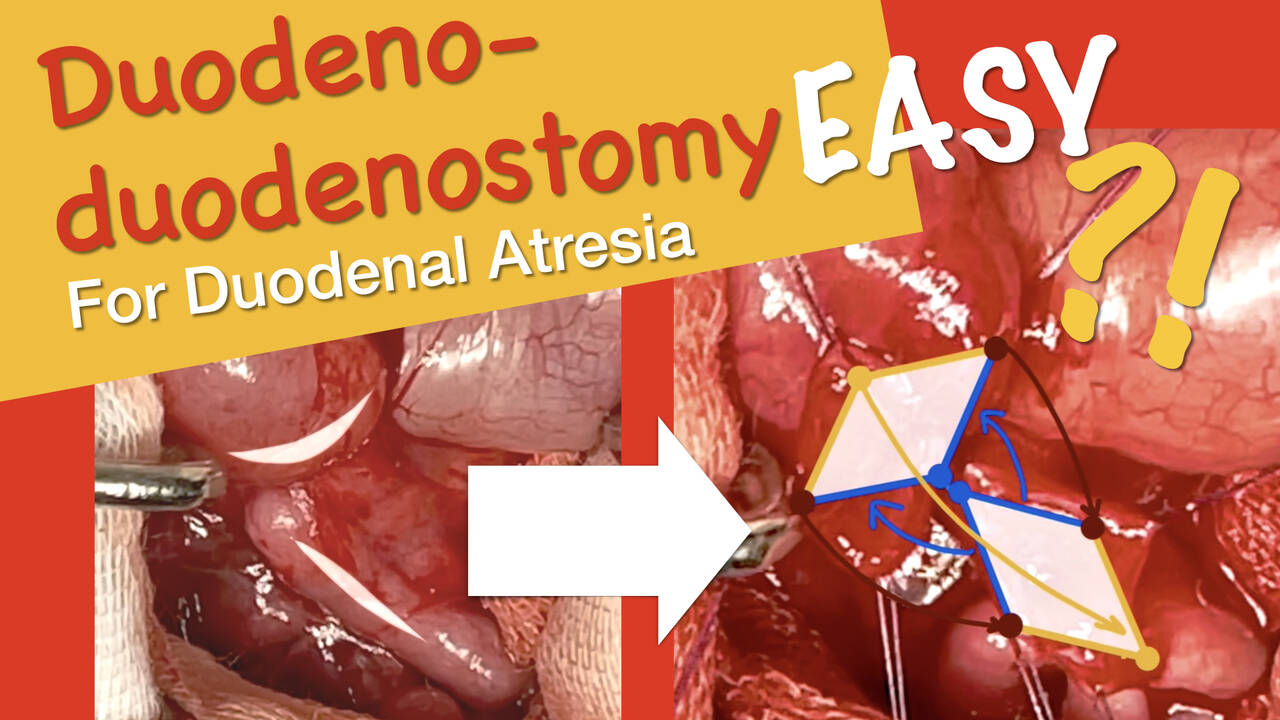
Duodeno-duodenostomy for Duodenal Atresia
7 min · Published Jun 2021
Video
Malrotation and Volvulus with Trinity
Dr. Steve Rothenberg · 8 min · Published May 2026
Video
Malrotation Rapid Fire Session: Update Course 2015
8 min · Published Jul 2017
Podcast
Malrotation with Dr. Meera Kotagal
13 min · Published May 2023
Video
Technique: Laparoscopic Repair of Duodenal Atresia, Ladd's Procedure, and...
Dr. Steve Rothenberg · 15 min · Published Nov 2018
Video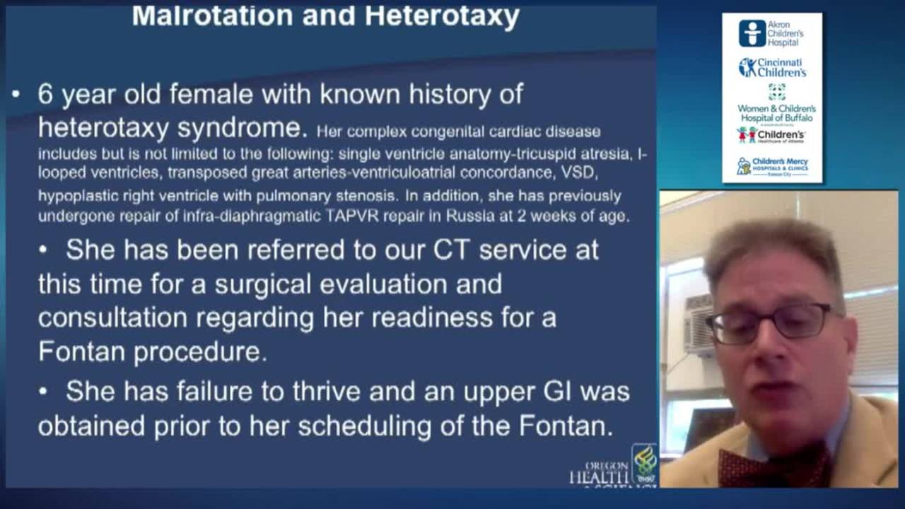
Malrotation - Volvulus - Meconium Ileus - Solid Organ Injury: Update Course 2015
27 min · Published Nov 2018
Video
Impact of social determinants of health on outcomes in pediatric short bowel syndrome...
43 s · Published Aug 2026
Video
Advancements in Pediatric Intestinal Failure: Innovative Therapies and Improved Outcomes
17 min · Published Aug 2026
Podcast
Update Course Rewind: Omphalocele & Gastroschisis 2020
Miguel Guelfand · 15 min · Published Jul 2026
Video
Update Course Rewind 2025: Updates in NEC Management
11 min · Published Jul 2026
Video
Availability, utilization, and barriers to bowel ultrasound for necrotizing enterocolitis...
55 s · Published Jul 2026
Video
Care transition from a pediatric intestinal rehabilitation program to adult care and the risk of all-cause mortality: A retrospective cohort study
1 min · Published May 2026
Video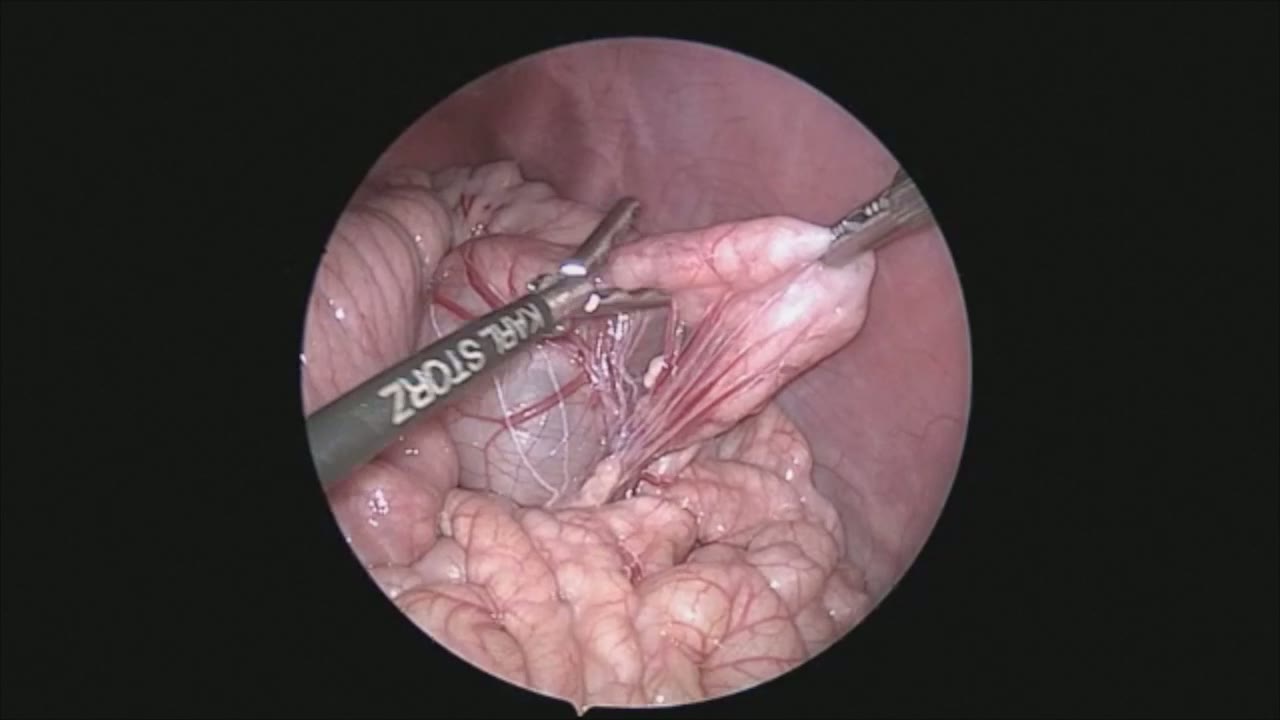
sealer demo
Dr. Steve Rothenberg · 3 min · Published May 2026
Video
pyloric knife
Dr. Steve Rothenberg · Published May 2026
Video
pulm artery LLL
Dr. Steve Rothenberg · 9 s · Published May 2026
Video
plication narrated
Dr. Steve Rothenberg · 6 min · Published May 2026
Video
percuvance redo nissen
Dr. Steve Rothenberg · 5 min · Published May 2026
Video
open bowel anast
Dr. Steve Rothenberg · Published May 2026
Video
Intestinal Atresia Types Explained: Grossfeld Classification for Pediatric Surgery
1 min · Published Sep 2026
Video
Derivation and validation of the Pediatric Community-Acquired Pneumonia Severity (PedCAPS) score: A prospective cohort study
50 s · Published Sep 2026
Video
FETO for Late-Diagnosed Severe Congenital Diaphragmatic Hernia (CDH) at Cincinnati Children's with Dr. Beth Rymeski
4 min · Published Sep 2026
Video
Severe Congenital Diaphragmatic Hernia (CDH) Case: FETO Management & Unexpected Findings with Dr. Beth Rymeski
3 min · Published Sep 2026
Video
Fetoscopic Endoluminal Tracheal Occlusion (FETO) Procedure: Step-by-Step Guide with Dr. Beth Rymeski
4 min · Published Sep 2026
Video
Choledochal Cyst Types Explained: Pediatric Surgery Fundamentals
1 min · Published Sep 2026
What the experts said
The 3 millimeter sealer can be used to safely grasp the bowel and act as a forceps which is atraumatic to the bowel.
The first portion of the duodenum is extremely dilated.
Dense adhesions to the proximal duodenum can be safely taken down using the sealer by dissecting them off the bowel and then sealing and gently tearing them off the surface of the duodenum.
Because of the minimal energy spread of the sealer, it is very safe to dissect in this fashion.
The bowel can be grasped immediately after the sealer is activated without any evidence of any heat injury to the bowel.
The sealer is found to be much more useful than using a hook, which had significant energy spread and also could not be used to grasp the bowel.
As is common in these cases, the duodenum goes towards the retroperitoneum, and this is the most difficult part to mobilize.
Adhesions between the transverse colon and the duodenum, some of which are consistent with Ladd bands, can be extremely thick and dense and difficult to take down.
Complete mobilization of the transverse and proximal or ascending colon is necessary because of the posterior attachments.
The minimal energy spread from the sealer allows this device to be used in close proximity to the small bowel without any risk of injury to it.
Ladd bands can traverse across the duodenum causing proximal obstruction.
The bowel is run from proximal to distal to completely derotate the bowel and eliminate the risk of volvulus in the future.
The posterior retroperitoneal attachment of the duodenum cannot be reached until the Ladd bands have been completely divided.
The sealer can act as an atraumatic bowel grasper during bowel running, though the jaws are not quite as large as the 3 millimeter bowel grasper.
The sealer is used during bowel running because other bands are often encountered, and this allows immediate sealing and division.
Enlarged lymph nodes in the mesentery and the chylous appearance within the bowel show evidence of chronic mild obstruction.
At the completion of the Ladd procedure, all of the colon is on the left and the small bowel is on the right.
In a small infant with a small appendix, the appendix can be brought out through the right trocar site and amputated extracorporeally.
The infant presented with repetitive bilious vomiting.
An upper GI series showed a redundant duodenum which did not cross the midline.
The surgeon is positioned at the end of the table with the baby brought down to the foot of the table to allow the surgeon to be in line with the foregut.
A 4 or 5 millimeter port is placed in the umbilicus, and right and left hand operating ports are placed either side of the umbilicus.
In a small infant, the right hand port is placed above the umbilicus so that the right hand does not conflict with the scope.