
Laparoscopic Jejunal Atresia Repair - Technique
With Dr. Dr. Steve Rothenberg · StayCurrentMD
Part of
Jejunal Atresia 3 items
Educational content from recorded physician discussions — not medical advice. Talk to your (or your child's) care team about your situation.
More about bowel obstruction
same diagnosisVideo
Laparoscopic Jejunal Atresia Repair - Technique
Dr. Steve Rothenberg · 5 min · Published Nov 2018
Podcast
Case-Based Journal Review- Intussusception in 2022
20 min · Published Feb 2022
Video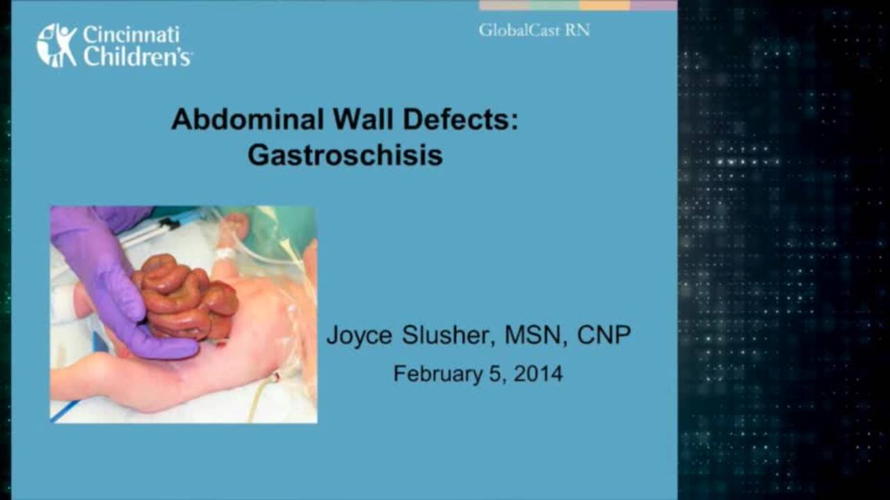
Gastroschisis: Advanced Practice Providers
43 min · Published Feb 2014
Podcast
Inguinal Hernias: Diagnosis and Management
16 min · Published Apr 2021
Video
Tricks - Laparoscopic Epigastric Herniorrhaphy
15 min · Published Nov 2018
Video
Challenges in Diaphragmatic Hernia Repair: Update Course 2016
44 min · Published Oct 2018
Only a few other public items share this expert — go deeper there →
Video
Pediatric Surgical Oncology Research Collaborative (PSORC): Studying Rare Pediatric Tumors
56 s · Published May 2026
Video
Update Course Rewind 2025: Hirschsprung + ARM: Rare but Real
1 min · Published May 2026
Video
Update Course Rewind 2025: Hirschsprung + ARM: Rare but Real
1 min · Published May 2026
Video
Pooling Patients to Study Rare Pediatric Tumors: An Introduction to PSORC
56 s · Published May 2026
Video
The fetal frontier: A review of current and emerging fetal therapies for genetic diseases
44 s · Published May 2026
Video
Indocyanine green assists with sentinel lymph node mapping in pediatric and adolescent patients
1 min · Published May 2026
What the experts said
A newborn with prenatal diagnosis of bowel obstruction was found to have jejunal atresia on upper GI study
The surgeon stands at the baby's feet with the cameraman on the baby's right and the scrub tech on the patient's left for this procedure
A 3-port technique was used with a 4 mm 30-degree scope for visualization and two 3 mm ports for dissection
The left mid-quadrant port was later changed to a 5 mm port for the stapler
The Veress needle is inserted below the umbilicus to avoid injury to the umbilical vessels and prevent CO2 embolism
There was a complete gap between the proximal jejunum and the distal bowel, with visualization of appendix and cecum suggesting an apple peel defect
The bowel was run from proximal to distal to ensure there were no other areas of obstruction or kinking
Running the bowel distally confirmed an apple peel defect, as the bowel could be seen twisting around the mesentery
Because there was no evidence of a kink or significant obstruction, the mesentery was not further manipulated
A decision was made to remove the proximal dilated segment in the hopes of improving bowel motility following surgery, because it was relatively short and significantly dilated
The mesentery is taken down using a 3 mm bipolar vessel sealer, with vessels clamped, sealed, and then teased off the mesenteric border of the bowel
The dissection was carried back almost to the ligament of Treitz and encompassed approximately 10 cm length of bowel
A 5 mm endoscopic stapler was used to divide the bowel, laying down 4 rows of staples and dividing between them
Two applications of the stapler were required because the bowel was so dilated, with a diameter of almost 4 cm
The distal jejunal segment was anastomosed to the proximal dilated segment in an end-to-side fashion
An enterotomy was made in the proximal dilated bowel using a 3 mm hook cautery and then decompressed with a 3 mm sucker
An upper GI obtained one week postoperatively showed a widely patent anastomosis
The resected specimen measured 10 cm
A similar enterotomy was made in the distal jejunal segment and slightly dilated to allow access of the stapler
The anastomosis was approximately 2.5 cm in length
The resultant enterotomy was closed with a running absorbable suture
Previous to having the stapler, an end-to-end anastomosis would have been performed with multiple interrupted or running sutures
After completion of the enterotomy, there was no evidence of significant mesenteric defect or any gap in the anatomy
The procedure took 80 minutes and was tolerated well by the infant
The patient had diminishing NG aspirates over the next week