


From
StayCurrentMD
QUAD #20: Endoscopic Repair of Minor Laryngeal Clefts with Dr. Catherine Hart
With Dr. Katherine Hart · hosted by Dr. Em Gootee
Chapter 1 of 5 · Fundamentals
Diagnostic challenge
Introduction and Diagnostic Challenge of Laryngeal Clefts
Expert statements on this page
No expert statements were drawn from this page.
Host summaries · secondary, not cited in answers
Educational content from recorded physician discussions — not medical advice. Talk to your (or your child's) care team about your situation.
Video
Laryngeal Clefts
CCHMC Pediatric Surgery · 14 min · Published Jun 2021
Video
Esophageal Atresia/Tracheoesophageal Fistula Bronchoscopy and Surgical Technique
Dr. Todd Ponsky · 16 min · Published Nov 2021
Video
QUAD #25: Medical Management of Minor Laryngeal Clefts with Dr. Greg Burg
CCHMC Pediatric Surgery · 7 min · Published Feb 2025
Video
Aerodigestive & Esophageal Surgery: Aspiration in TEFs
5 min · Published Dec 2014
Video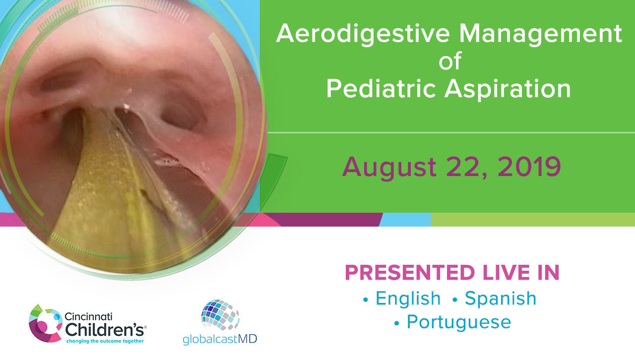
Aerodigestive Management of Pediatric Aspiration - FULL SHOW
CCHMC Pediatric Surgery · 157 min · Published Jul 2020
Video
2025 Pediatric Surgery Update Course - Updates in Esophageal Atresia Management
16 min · Published Aug 2025
Video
Surgeon annual volume impacts recurrence rates of pediatric inguinal hernia repairs: A multi-institutional study
54 s · Published Feb 2026
Podcast
Journal of Pediatric Surgery Article Review: 2nd Quarter (Apr-Jun) 2025
16 min · Published Aug 2025
Video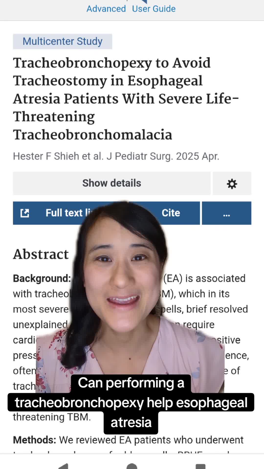
Tracheobronchopexy to Avoid Tracheostomy in Esophageal Atresia Patients With Severe Life-Threatening Tracheobronchomalacia
50 s · Published Apr 2025
Video
QUAD #27 - Multidisciplinary - How Do Teams Enhance Outcomes by the CCHMC ADEC Team
CCHMC Pediatric Surgery · 10 min · Published Apr 2025
Video
QUAD #22: What is CHARGE syndrome? with Dr. Catherine Hart
CCHMC Pediatric Surgery · 59 s · Published Dec 2024
Video
QUAD #5 Pharyngeal Scar Management with Dr. Doug von Allmen
CCHMC Pediatric Surgery · 7 min · Published Mar 2024
Video
Intestinal Atresia Types Explained: Grossfeld Classification for Pediatric Surgery
1 min · Published Sep 2026
Video
Derivation and validation of the Pediatric Community-Acquired Pneumonia Severity (PedCAPS) score: A prospective cohort study
50 s · Published Sep 2026
Video
FETO for Late-Diagnosed Severe Congenital Diaphragmatic Hernia (CDH) at Cincinnati Children's with Dr. Beth Rymeski
4 min · Published Sep 2026
Video
Severe Congenital Diaphragmatic Hernia (CDH) Case: FETO Management & Unexpected Findings with Dr. Beth Rymeski
3 min · Published Sep 2026
Video
Fetoscopic Endoluminal Tracheal Occlusion (FETO) Procedure: Step-by-Step Guide with Dr. Beth Rymeski
4 min · Published Sep 2026
Video
Choledochal Cyst Types Explained: Pediatric Surgery Fundamentals
1 min · Published Sep 2026
What the experts said
A type 1 laryngeal cleft can appear normal on flexible bronchoscopy but be visible on rigid bronchoscopy using less sophisticated identification techniques.
Combined flexible and rigid bronchoscopy evaluations are complementary and necessary to avoid missing laryngeal clefts.
Surgical indications for laryngeal cleft repair include ongoing respiratory symptoms, aspiration, failure to thrive, recurrent pulmonary infections, and failure of conservative measures (or in some instances before trying conservative measures).
Mass closure or layered closure technique can be used for laryngeal cleft repair, but the key requirement is that edges must be inverted when finished.
Cold steel is preferred over laser for minor cleft resection because it is simpler to set up, faster, and eliminates the risk of laser fire.
Closure is performed using interrupted sutures, typically PDS on an RB1 or P3 needle depending on child size.
Postoperatively, children are observed overnight on the airway unit and resume their preoperative diet, which is continued until postoperative evaluation.
Postoperative evaluation is typically performed 6 to 8 weeks after surgery and includes repeat video swallow study and repeat endoscopy to ensure healing.
Failure of endoscopic cleft repair is not super common.
Incomplete demucosalization at the apex can result in healing at the top but a gap remaining at the back.
Layered closure technique (closing anterior and posterior portions separately) seems like more effort in minor clefts but is a more reasonable option for type 2 or 3 clefts to ensure good closure.
Conservative management (not surgery) achieves resolution over time in 51% of children with laryngeal clefts.
Injection laryngoplasty achieves improvement in symptoms in two-thirds of children and resolution in one-third.
Complications of surgical cleft repair are often not terrible but include reports of laryngeal scarring, supraglottic infections, and lacerations.
The goal of endoscopic cleft repair is to remove the interarytenoid mucosa to create two raw surfaces and then suture the edges together.
If suture bites are taken too deeply during cleft repair, edges will be inverted and the child will continue to have problems.
Interarytenoid mucosa can be resected using laser or cold steel (knife), and the choice does not matter clinically.
Complete demucosalization of the apex is essential; if incomplete, a hole will remain at the apex and the child will continue to aspirate.
Typically 2 to 3 sutures are placed depending on the extent of the cleft.
Epiglottic folds are released after suturing to create extra space.
Surgical closure of laryngeal clefts achieves improvement in symptoms in almost 80% of children and resolution in 70%.
Surgical closure has a slightly higher risk of complications compared to conservative management.
A consensus guideline exists for diagnosing and managing laryngeal clefts in children and is a good resource, especially for those newer to practice.