


From
StayCurrentMD
Technique: Laparoscopic Repair of Duodenal Atresia, Ladd's Procedure, and...
With Dr. Steve Rothenberg
Chapter 1 of 4 · Surgical Management
Setup & ports
Room Setup, Port Placement, and Initial Dissection
Expert statements on this page
No expert statements were drawn from this page.
Host summaries · secondary, not cited in answers
Educational content from recorded physician discussions — not medical advice. Talk to your (or your child's) care team about your situation.
Video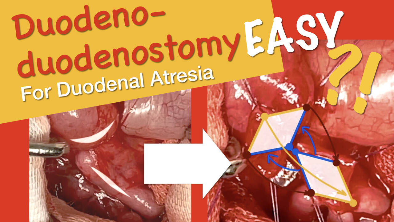
Duodeno-duodenostomy for Duodenal Atresia
7 min · Published Jun 2021
Video
Peritoneal Access
Published Mar 2020
Podcast
Intestinal Rehabilitation, Episode 2: Overwhelming intestinal damage, Part 1
13 min · Published Feb 2022
Video
Enhanced Recovery After Surgery (ERAS) Program: Update Course 2016
44 min · Published Jul 2017
Video
Antibiotic Stewardship - APSA Practice Gaps 2019
Published Mar 2020
Video
BOB in Ped Surg 2023 - CIPESUR Winner - Georgina Falcioni, MD
Published Feb 2023
Video
sealer demo
Dr. Steve Rothenberg · 3 min · Published May 2026
Video
pyloric knife
Dr. Steve Rothenberg · Published May 2026
Video
pulm artery LLL
Dr. Steve Rothenberg · 9 s · Published May 2026
Video
plication narrated
Dr. Steve Rothenberg · 6 min · Published May 2026
Video
percuvance redo nissen
Dr. Steve Rothenberg · 5 min · Published May 2026
Video
open bowel anast
Dr. Steve Rothenberg · Published May 2026
Video
Intestinal Atresia Types Explained: Grossfeld Classification for Pediatric Surgery
1 min · Published Sep 2026
Video
Derivation and validation of the Pediatric Community-Acquired Pneumonia Severity (PedCAPS) score: A prospective cohort study
50 s · Published Sep 2026
Video
FETO for Late-Diagnosed Severe Congenital Diaphragmatic Hernia (CDH) at Cincinnati Children's with Dr. Beth Rymeski
4 min · Published Sep 2026
Video
Severe Congenital Diaphragmatic Hernia (CDH) Case: FETO Management & Unexpected Findings with Dr. Beth Rymeski
3 min · Published Sep 2026
Video
Fetoscopic Endoluminal Tracheal Occlusion (FETO) Procedure: Step-by-Step Guide with Dr. Beth Rymeski
4 min · Published Sep 2026
Video
Choledochal Cyst Types Explained: Pediatric Surgery Fundamentals
1 min · Published Sep 2026
What the experts said
The patient is a 1 day old infant who had a prenatal diagnosis of duodenal atresia
Port placements consist of a 4 millimeter scope in the umbilicus, a 3 millimeter port in the left mid quadrant, and a 3 millimeter stab wound in the right mid quadrant, triangulated towards the right upper quadrant
The 3 millimeter sealer is an excellent dissecting device and can be used in safe energy in close approximation to both the gallbladder and the small bowel
A transabdominal stitch using a 30 Prolene on an RB1 needle is placed through the anterior abdominal wall in the right upper quadrant to retract the gallbladder superiorly
The child is malrotated with a complete duodenal atresia
The bowel is run from proximal to distally to ensure that there are no further Ladd's bands and to completely de-rotate the bowel
A Meckel's diverticulum is found in the distal ileum during the procedure
All of the small bowel is placed on the patient's right and the large bowel on the patient's left
Widening the mesenteric base should diminish the chance of volvulus in the future
A transverse enterotomy is made at the most dependent portion of the dilated proximal segment
The enterotomy in the distal segment is longitudinal
There is increased smoke and debris from using electrocautery compared to the 3 millimeter sealer
A transabdominal stitch aligns the two sides of the bowel to perform the side to side anastomosis
A pre-cut Vicryl suture approximately 10 to 12 centimeters in length is used to run the back row of the anastomosis
In some cases, the distal duodenal segment is more fixed to the retroperitoneum, making this part of the procedure more difficult, but the same technique is used with excellent result
The surgeon prefers running suture over interrupted suture as it works extremely well and is more efficient
The entire procedure took approximately 50 minutes
Approximately 10% of patients undergoing laparoscopic repair of duodenal atresia have malrotation
The patient is a 2.8 kg child
A nasogastric tube is left to suction for approximately 3 to 4 days, and in most cases feeds are started by the 5th day
This child started eating on the 5th postoperative day and was on full feeds by the 8th postoperative day
The base of the Meckel's diverticulum could adequately be divided without compromising the lumen of the bowel, so a decision was made to remove it using the 5 millimeter stapler
The left quadrant 3 millimeter port was changed to a 5 millimeter port to allow insertion of the endoscopic stapler
Because of the malrotation, the appendix was also removed
The child was discharged to home on the 10th postoperative day