
 StayCurrentMD
Duodenal Atresia
Everything in the library about duodenal atresia — built automatically from the recorded discussions that name it
Educational content from recorded physician discussions — not medical advice. Always talk to your child's care team about your child's situation.
Content of this collection
Surgical Management
2 items
Technique: Laparoscopic Repair of Duodenal Atresia, Ladd's Procedure, and...
Watch →
Laparoscopic Repair of Duodenal Atresia, Ladd's Procedure, and Meckel's Diverticulectomy in a Newborn using Just Right stapler by Dr. Steven Rothenberg.
video15:01 · Nov 2018
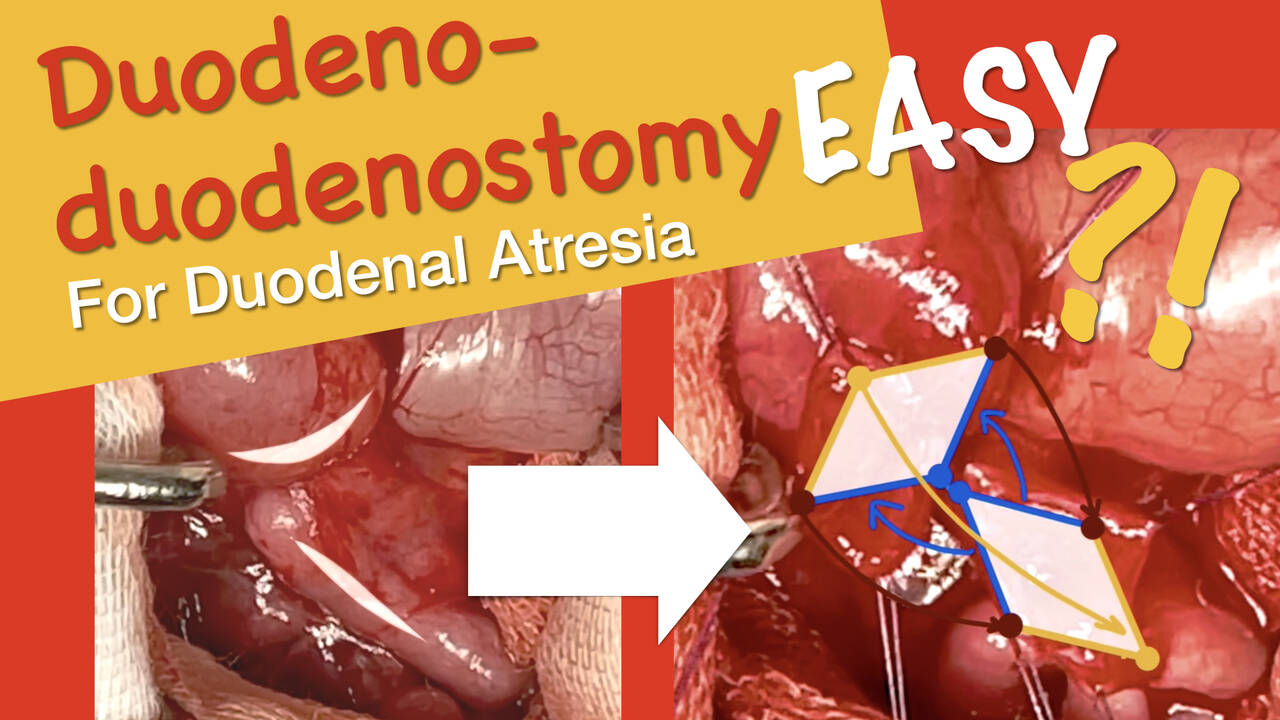
Duodeno-duodenostomy for Duodenal Atresia
Watch →
In this video, duodeno-dudenostomy diamond anastomsis for duodenal atresia is demonstrated. The patient is a 2 day old boy that is presented with greenish vomiting since birth. Exploration revealed complete duodenal atresia type three and m
video7:04 · Jun 2021
Evidence & Research
1 item

BOB in Ped Surg 2023 - CIPESUR Winner - Georgina Falcioni, MD
Watch →
Watch the CIPESUR winner, Georgina Falcioni, MD, present her presentation on "Comparative effectiveness of Telesimulation vs standard simulation for MIS essential skills training."
video · Feb 2023
Summaries and takeaways+ Show
The doctors in this collection+ Show
All expert statements+ Show
Every expert statement below comes from the recorded discussions, with its speaker and moment.
Duodeno-duodenostomy for Duodenal Atresia
The patient is a two-day-old boy with duodenal atresia.
host_summary0:00 ↗
A transverse right abdominal incision allows access to the duodenum and small intestine.
host_summary0:20 ↗
Malrotation is present in this case.
host_summary1:00 ↗
Duodenal atresia type III is present in this case.
host_summary1:00 ↗
The proximal duodenal opening is made at the most dependent part.
host_summary2:00 ↗
The distal limb is opened on the anterolateral surface to avoid injury to the opening of the common bile and pancreatic ducts.
host_summary2:30 ↗
Bile coming out on opening of the distal limb confirms patency to that point.
host_summary2:45 ↗
The diamond-shaped technique creates two openings with perpendicular axes.
host_summary2:55 ↗
Distal patency is checked by injecting saline and observing its filling to the whole bowel.
host_summary3:10 ↗
The anastomosis technique uses two opposing borders of the diamonds to make the posterior wall and the far-facing borders to make the anterior wall.
host_summary3:25 ↗
The posterior wall is sutured from the inside with full-thickness sutures.
host_summary3:50 ↗
The anterior wall is sutured from the outside.
host_summary4:00 ↗
5-0 absorbable sutures are used for the anastomosis.
host_summary4:10 ↗
Suturing starts with the middle of the posterior wall and advances toward the angles with interrupted sutures.
host_summary4:10 ↗
The angle suture is placed from the outside in an extramucosal fashion.
host_summary4:35 ↗
The anterior wall sutures are placed in an extramucosal fashion starting from the angles and working towards the middle.
host_summary5:10 ↗
The proximal dilated pouch is brought downwards to meet the distal limb during anterior wall closure.
host_summary5:30 ↗
Widening of the root of the mesentery is performed after completing the anastomosis.
host_summary5:50 ↗
The superior mesenteric vessels should be visualized during mesenteric root widening.
host_summary6:05 ↗
The peritoneal covering is carefully dissected to allow adequate widening of the mesenteric root.
host_summary6:15 ↗
Appendectomy is performed as part of the procedure.
host_summary6:30 ↗
The bowel is returned to the abdomen with the colon in the left side and the duodenojejunal junction in a straight direction.
host_summary6:40 ↗
Technique: Laparoscopic Repair of Duodenal Atresia, Ladd's Procedure, and...
The patient is a 1 day old infant who had a prenatal diagnosis of duodenal atresia
clinical0:00 ↗
Port placements consist of a 4 millimeter scope in the umbilicus, a 3 millimeter port in the left mid quadrant, and a 3 millimeter stab wound in the right mid quadrant, triangulated towards the right upper quadrant
clinical0:30 ↗
The 3 millimeter sealer is an excellent dissecting device and can be used in safe energy in close approximation to both the gallbladder and the small bowel
opinion1:00 ↗
A transabdominal stitch using a 30 Prolene on an RB1 needle is placed through the anterior abdominal wall in the right upper quadrant to retract the gallbladder superiorly
clinical2:00 ↗
The child is malrotated with a complete duodenal atresia
clinical2:30 ↗
The bowel is run from proximal to distally to ensure that there are no further Ladd's bands and to completely de-rotate the bowel
clinical3:00 ↗
A Meckel's diverticulum is found in the distal ileum during the procedure
clinical3:30 ↗
All of the small bowel is placed on the patient's right and the large bowel on the patient's left
clinical4:00 ↗
What's new+ Show