


Chapter 1 of 6 · Diagnosis & Workup
Technique overview
Contrast enema technique for Hirschsprung diagnosis
Expert statements on this page
No expert statements were drawn from this page.
Host summaries · secondary, not cited in answers
Educational content from recorded physician discussions — not medical advice. Talk to your (or your child's) care team about your situation.
Video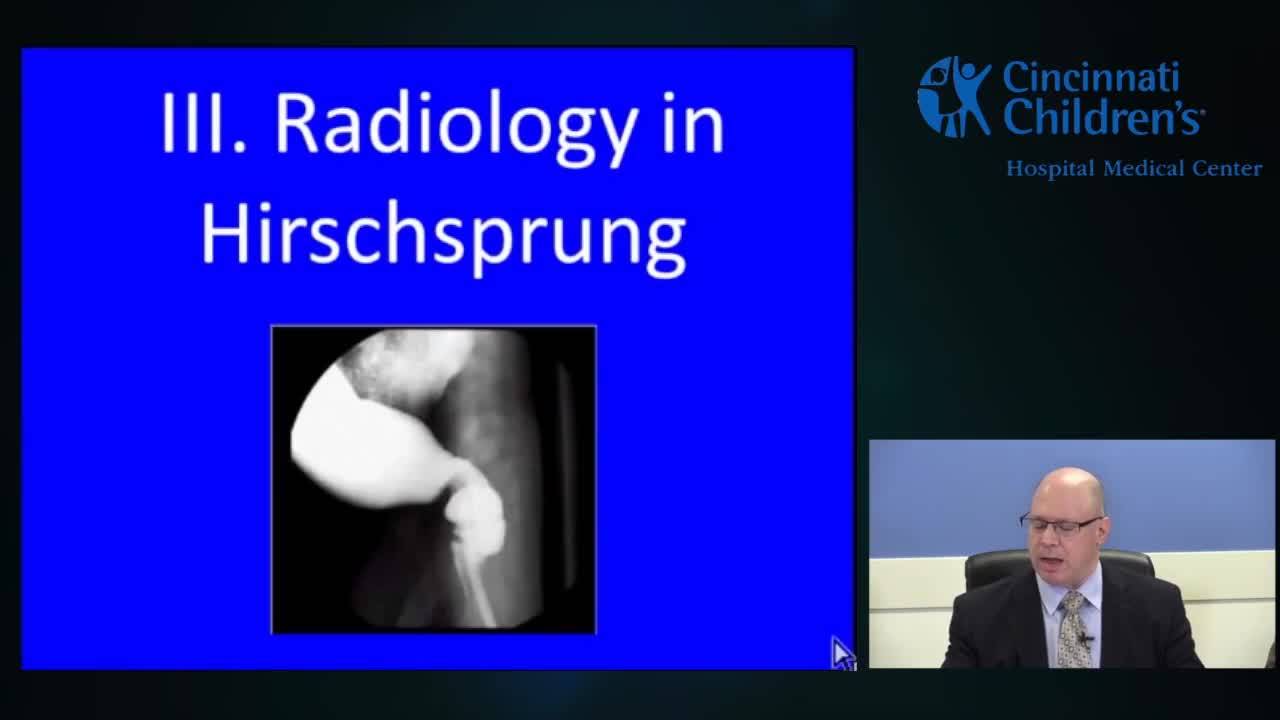
Evaluation & Management Of Hirschsprung's Disease
44 min · Published Nov 2018
Video
Hirschsprung Disease: Radiology Aspect
CCHMC Pediatric Surgery · 43 min · Published Feb 2015
Video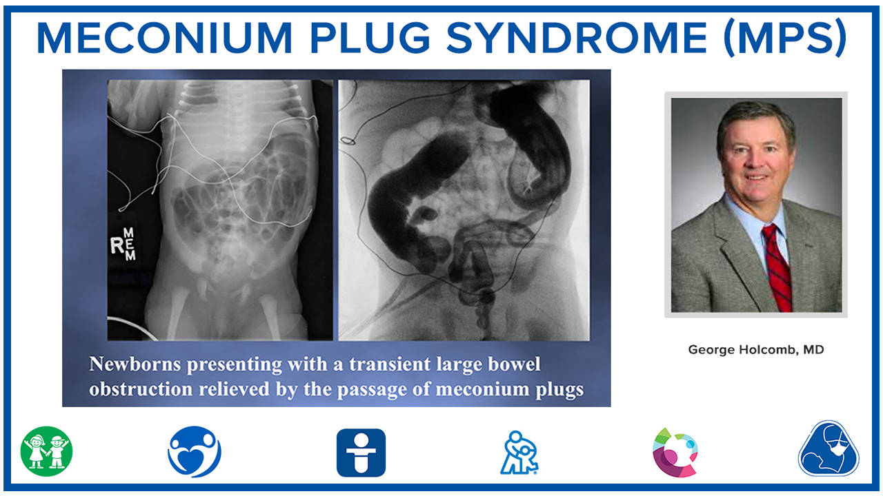
Meconium Plug Syndrome (MPS)
Published Mar 2020
Podcast
The Colorectal Quiz Episode 10: Total Colonic Hirschsprung Disease Part 1
12 min · Published Apr 2021
Podcast
Colorectal Quiz Episode 3: Hirschsprung Disease
20 min · Published Feb 2021
Video
Hirschsprung Disease: Update Course 2015
CCHMC Pediatric Surgery · 7 min · Published Nov 2015
Video
Validation of an anorectal malformation trainer - Can a high-fidelity model simulate real life?
1 min · Published Jun 2026
Video
Complications and Long-Term Outcomes of Patients With Cloacal Malformation After Bowel Neovagina...
1 min · Published Jun 2026
Video
Safety and utility of long-acting steroid injection for management of post-operative stricture...
1 min · Published Jun 2026
Video
Association Between Social Determinants of Health and Choice of Urinary Reconstruction in Children
1 min · Published Jun 2026
Video
The Perineal Body Preserving PSARP (PPP)
11 min · Published Jun 2026
Video
Update Course Rewind 2025: Hirschsprung + ARM: Rare but Real
1 min · Published May 2026
Video
Radiology and Image Diagnosis of Hirschsprung Disease
44 min · Published Nov 2018
Video
Outcomes and Complications in Hirschsprung Disease
105 min · Published Nov 2018
Video
Surgical Procedures for Hirschsprung Disease
128 min · Published Nov 2018
Video
Cloaca - Prental Imaging & Diagnosis - Counseling
Dr. Todd Ponsky · 36 min · Published Nov 2018
Podcast
Cloacal Exstrophy with Dr. Alberto Peña
53 min · Published Apr 2017
Video
Hirschsprung Disease: Surgical Procedures
128 min · Published Feb 2015
Video
Intestinal Atresia Types Explained: Grossfeld Classification for Pediatric Surgery
1 min · Published Sep 2026
Video
Derivation and validation of the Pediatric Community-Acquired Pneumonia Severity (PedCAPS) score: A prospective cohort study
50 s · Published Sep 2026
Video
FETO for Late-Diagnosed Severe Congenital Diaphragmatic Hernia (CDH) at Cincinnati Children's with Dr. Beth Rymeski
4 min · Published Sep 2026
Video
Severe Congenital Diaphragmatic Hernia (CDH) Case: FETO Management & Unexpected Findings with Dr. Beth Rymeski
3 min · Published Sep 2026
Video
Fetoscopic Endoluminal Tracheal Occlusion (FETO) Procedure: Step-by-Step Guide with Dr. Beth Rymeski
4 min · Published Sep 2026
Video
Choledochal Cyst Types Explained: Pediatric Surgery Fundamentals
1 min · Published Sep 2026
What the experts said
The contrast agent used is iodinated, water-soluble, and hyperosmotic with osmolality approximately 400.
The hyperosmotic contrast (osmolality ~400) is similar to agents used for colon cleansing and can help clean the colon in addition to making the diagnosis.
In neonates, retained hyperosmotic contrast can cause dehydration and clinical deterioration, requiring neonatal ICU awareness.
Gravity infusion is used rather than injection, with moderate-pace infusion to rapidly visualize distal and proximal segments and identify transition zones.
Lateral rectosigmoid imaging is performed to visualize the transition zone.
Early maximal distention is best for seeing the transition zone; delayed imaging can cause distention of the distal aganglionic segment because it is soft tissue, not a rigid lead pipe.
If the colon appears small in a neonate, the entire colon is filled with attempt to reflux into terminal ileum to identify other diagnoses.
In full-term neonates, a 12-14 French Foley catheter is used; in premature infants, a smaller size is used.
On normal contrast enema, the rectum is well distended, presacral space is well seen on true lateral image (femurs superimposed), and proximal colon toward splenic flexure is slightly smaller than rectum.
Very short-segment Hirschsprung disease can be missed if a Foley balloon is inflated in the distal rectum, blocking visualization of the transition zone.
Rectosigmoid transition zone Hirschsprung cases are usually concordant between radiologic and pathologic findings.
In long-segment Hirschsprung disease, the rectum is very small and there is irregular, spastic-appearing colon extending to the splenic flexure or beyond.
In total colonic Hirschsprung disease, the rectum is not larger than the rest of the colon, and the entire colon appears uniformly small.
In premature infants, the contrast enema does not follow the diagnostic rules because the colon may appear immature and small, making it impossible to distinguish from pathology.
Contrast enema can be performed in premature infants as young as 35-36 weeks gestational age with reasonable diagnostic accuracy.
Below 35-36 weeks gestational age, especially in the setting of necrotizing enterocolitis, diagnostic accuracy of contrast enema is reduced.
Rectosigmoid transition in Hirschsprung disease is located at approximately the S2 vertebral level; distal rectal disease is below S1-S2.
Small left colon syndrome typically has a transition at the splenic flexure that is very abrupt.
The rectosigmoid index (rectum larger than sigmoid) is a useful principle but not definitive; imaging must extend to the splenic flexure to avoid missing proximal disease.
A case initially interpreted as small left colon (small rectum, small colon to splenic flexure, meconium plugs) was proven to be total colonic aganglionosis with terminal ileum transition.
When the transition zone appears proximal (splenic flexure or beyond), the radiologic transition zone cannot accurately predict the histologic transition zone.
Proximal transition zones should prompt consideration of more invasive surgical approaches (laparoscopic or open) rather than transanal pull-through, because the true transition may be much more proximal than radiologically apparent.
One panelist performs rectal biopsy in almost any patient requiring contrast enema to rule out distal obstruction, including meconium plug, small left colon, and meconium ileus.
One panelist does not perform rectal biopsy in clear cases of meconium ileus with terminal ileum reflux and clinical improvement.
Dr. Pena would not perform rectal biopsy if certain the diagnosis is meconium ileus, but would perform biopsy for small left colon because it cannot be reliably distinguished from Hirschsprung disease radiologically.