
Collection
Abdominal Compartment Syndrome
Educational content from recorded physician discussions — not medical advice. Always talk to your child's care team about your child's situation.
Content of this collection
Resources
8 items

Approach and component separation for suture closure and underlay mesh...
Watch →
During the Pediatric Surgery Tricks of the Trade and Difficult Cases: Innovative Solutions to Common Problems Course in 2013, directors DrsTodd Ponsky, Robert Parry, and Jacob Langer, along with faculty including Drs David van der Zee, Suad
video31:03 · Sep 2018
Abdominal Wall Defects
Watch →
Dr. Jacob Langer discusses various abdominal wall defects through case presentation.Topics of discussion include atresia, gastroschisis, fascial defects, staged closure by silo, and ruptured omphalocele.
video37:50 · Nov 2018
Gastroschisis: Advanced Practice Providers
Watch →
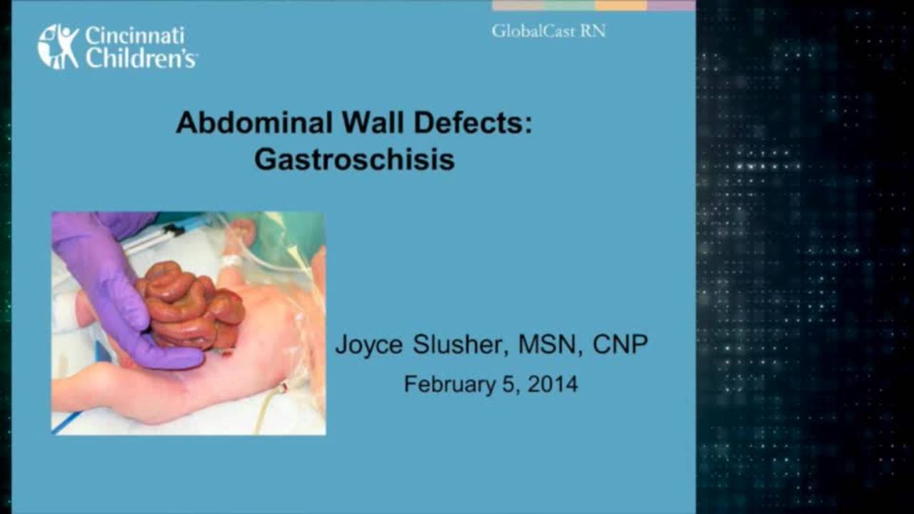
Joyce Slusher, MSN, CNP, presents on perioperative management of gastroschisis for advanced practice providers.
video43:21 · Jan 2019
Abdominal Wall Defects: Update Course 2013
Watch →
Dr. Jacob Langer discusses various abdominal wall defects through case presentation.Topics of discussion include atresia, gastroschisis, fascial defects, staged closure by silo, and ruptured omphalocele.
video37:40 · Jan 2019
Staged Closure of Gastroschisis with Spring-loaded Silo
Watch →
This video appears in a new pediatric surgery textbook, Clinical Pediatric Surgery: A Case-Based Interactive Approach," by Dr. Sherif Emil. The book is an innovative educational resource that focuses on judgment and decision-making in pedia
video27:11 · Feb 2020
Update Course 2022 - TOP PUBLICATIONS IN NON- PED SURG JOURNALS - Jose Campos
Watch →
In this session, Dr. Jose Campos leads us through the top publications that are not in the Journal of Pediatric Surgery.
Articles referenced:
1. IMPPACT (Intravenous Monotherapy for Postoperative Perforated Appendicitis in Children T
video27:50 · Sep 2022
Use of a new vertical traction device for early traction-assisted staged closure of congenital abdominal wall defects: a prospective series of 16 patients
Watch →
Anna-Maria Ziegler, Daniel Svoboda, Britta Lüken-Darius, Andreas Heydweiller, Fritz Kahl, Sophie Christine Falk, Udo Rolle, Till-Martin Theilen
Purpose: Abdominal wall closure in patients with giant omphalocele (GOC) and complicated gast
video0:56 · Nov 2024
Quick Literature Updates Episode 20
Watch →
We’re back with 20th episode of "Quick Literature Updates" the podcast series that delivers the latest updates in pediatric surgery literature in a quick and digestible format. In each episode, we review articles covering the most interesti
video4:03 · Jun 2025
Summaries and takeaways+ Show
Summary of this collection+ Show
Key points, with the moment each was said+ Show
Featured picks for families are being prepared — the summaries above are written for you.
Takeaways+ Show
Abdominal compartment syndrome is a critical concern in the management of congenital abdominal wall defects, particularly giant omphalocele and gastroschisis. Monitoring intra-abdominal pressure during and after closure is essential, with ventilatory parameters serving as the most reliable measure of safe closure tension [e639-c33, e1059-c24]. Bladder pressure measurement, though standard practice at some centers for 24 hours post-closure, has questioned reliability in tiny newborns [e639-c34, e1059-c23]. Intragastric pressure via NG tube offers an easy intraoperative guide, with 20 mmHg suggested as a threshold; pressures of 12 mmHg with a stable baby provide reassurance, while 35-40 mmHg raises concern even if the baby appears stable [e639-c35, e639-c36, e1059-c22]. Complications of primary gastroschisis closure stem mainly from increased abdominal pressure leading to decreased venous return and possible abdominal compartment syndrome with ischemic injury . In older children with giant omphalocele, rapid reduction can cause abdominal compartment syndrome and death; time must be allowed for abdominal domain expansion, with staged enlargement of the fascial defect over 2-3 stages preventing forced reduction [e639-c39, e1059-c25, e1059-c26]. Novel traction-assisted closure devices have shown promise: complete fascial closure was achieved after a median of 7 days in giant omphalocele and 5 days in complicated gastroschisis, with no patients developing abdominal compartment syndrome [e9436-c3, e9436-c4, e9436-c5, e10554-c6, e10554-c7, e10554-c8].
- Ventilatory parameters are the most reliable measure for assessing safe closure tension; bladder pressures in neonates are unreliable.
- Intragastric pressure monitoring via NG tube is easy intraoperatively; 20 mmHg is a suggested threshold, with 35-40 mmHg raising concern.
- Primary gastroschisis closure complications stem from increased abdominal pressure causing decreased venous return and possible compartment syndrome with ischemic injury.
- In older children with giant omphalocele, rapid reduction risks compartment syndrome and death; staged enlargement over 2-3 sessions prevents forced reduction.
- Traction-assisted closure devices achieve fascial closure in 5-7 days for gastroschisis and giant omphalocele without compartment syndrome or ventral hernias at 12 months.
For patients & families
Abdominal compartment syndrome is a serious complication that can occur when doctors are repairing birth defects of the abdominal wall, such as gastroschisis (where intestines are outside the body through a hole) or omphalocele (where organs are covered by a sac outside the body) [e1049-c27, e639-c39]. When surgeons try to put the organs back inside and close the abdomen, sometimes the pressure inside becomes too high, which can squeeze blood vessels and make it hard for the baby to breathe [e1049-c27, e1049-c28]. To prevent this dangerous situation, doctors carefully monitor several measurements during and after surgery: they watch the baby's breathing pressures on the ventilator, check urine output, examine the belly for tightness, and sometimes measure pressure inside the bladder or stomach [e1049-c28, e639-c33, e639-c36, e1059-c22, e1059-c24]. If the pressure gets too high during closure, surgeons will stop and use a temporary covering called a silo to allow gradual reduction over several days instead [e430-c39, e1049-c21, e2251-c4]. In older children with large defects, doctors must work even more slowly because their abdomens don't expand as easily as newborns', and rushing closure could cause compartment syndrome and be life-threatening [e639-c39, e1059-c25, e1059-c26]. New devices that apply gentle traction over several days have helped achieve safe closure without causing compartment syndrome .
Abdominal compartment syndrome is a serious complication that can occur when doctors are repairing birth defects of the abdominal wall, such as gastroschisis (where intestines are outside the body through a hole) or omphalocele (where organs are covered by a sac outside the body) [e1049-c27, e639-c39]. When surgeons try to put the organs back inside and close the abdomen, sometimes the pressure inside becomes too high, which can squeeze blood vessels and make it hard for the baby to breathe [e1049-c27, e1049-c28]. To prevent this dangerous situation, doctors carefully monitor several measurements during and after surgery: they watch the baby's breathing pressures on the ventilator, check urine output, examine the belly for tightness, and sometimes measure pressure inside the bladder or stomach [e1049-c28, e639-c33, e639-c36, e1059-c22, e1059-c24]. If the pressure gets too high during closure, surgeons will stop and use a temporary covering called a silo to allow gradual reduction over several days instead [e430-c39, e1049-c21, e2251-c4]. In older children with large defects, doctors must work even more slowly because their abdomens don't expand as easily as newborns', and rushing closure could cause compartment syndrome and be life-threatening [e639-c39, e1059-c25, e1059-c26]. New devices that apply gentle traction over several days have helped achieve safe closure without causing compartment syndrome .
The doctors in this collection+ Show
All expert statements+ Show
Every expert statement below comes from the recorded discussions, with its speaker and moment.
Abdominal Wall Defects
Most damage to the intestine in gastroschisis occurs in the last few weeks of pregnancy, supported by animal and clinical studies.
clinical2:45 ↗
Delivering gastroschisis at 37 weeks results in better neonatal outcomes compared to waiting for spontaneous labor.
clinical3:14 ↗
The age at spontaneous onset of labor for gastroschisis pregnancy is 3 weeks earlier than the general population.
epidemiological3:44 ↗
About half of gastroschisis pregnancies go into labor spontaneously before 37 weeks.
epidemiological3:56 ↗
No perinatal center in Canada performs routine cesarean sections for gastroschisis.
epidemiological4:37 ↗
Routine cesarean section for gastroschisis was standard of care 15-20 years ago but has gone out of favor.
guideline4:49 ↗
Using forceps and retractors for bedside reduction can harm the bowel in some cases.
clinical8:18 ↗
A pre-formed silo allows gentle reduction of gastroschisis contents without harming the bowel, and in about one-third of cases the bowel can be reduced immediately and the silo removed.
clinical8:27 ↗
Waiting for the neonate to lose 10% of body weight reduces bowel edema and makes reduction easier.
clinical9:14 ↗
Leaving a silo on for more than one day causes the fascial defect to enlarge significantly.
clinical9:41 ↗
A larger fascial defect after silo removal takes longer to contract and heal if not surgically closed.
clinical9:54 ↗
Rafensberger's group closed 80% of gastroschisis cases primarily; current practice has decreased to one-third to one-half primary closures.
epidemiological10:02 ↗
Plastic closure (non-surgical closure with dressing) produces extremely good results for gastroschisis, often with a small umbilical hernia that closes by age 2 in the vast majority of cases.
clinical11:37 ↗
Plastic closure avoids the need for operating room and general anesthesia in successful bedside reductions.
clinical11:50 ↗
There are two types of intestinal atresia in gastroschisis: early-developing atresia not always associated with bowel thickening, and late atresia due to very small abdominal wall defect.
clinical15:58 ↗
Primary repair of atresia in gastroschisis is appropriate when the bowel does not look damaged.
clinical15:00 ↗
If the bowel looks nasty or matted, the atresia should be managed by reduction and delayed repair at 6 weeks.
clinical15:09 ↗
Bringing an ostomy out through the umbilicus avoids a lateral scar and makes appliance placement easier.
clinical16:41 ↗
Gord Cameron first described umbilical ostomies in the 1980s.
epidemiological17:53 ↗
At 4 weeks post-gastroschisis repair with feeding intolerance, waiting longer is reasonable; at 8 weeks, exploration is warranted.
opinion20:07 ↗
A mechanical stricture causing feeding intolerance after gastroschisis repair, when fixed surgically, can result in full feeds within 1-2 weeks.
clinical19:46 ↗
Removing the omphalocele sac and attempting closure can lead to inability to achieve closure and need for prosthetic patch.
clinical22:13 ↗
A partially ruptured omphalocele sac can be closed and painted, functioning as an autogenous silo.
clinical22:23 ↗
Schuster-type repair uses mesh sutured to fascia with gradual closure over the intact sac, eventually allowing primary repair.
clinical22:31 ↗
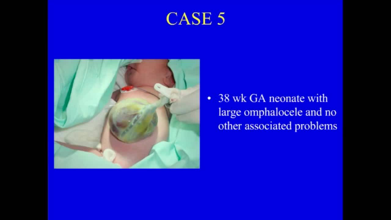
For large omphalocele in a full-term baby with no other problems, staged closure with patch and skin coverage is preferable to paint-and-wait, which takes months.
opinion23:05 ↗
Lateral component separation can facilitate bringing fascial edges together in omphalocele closure.
clinical23:32 ↗
Keeping the omphalocele sac supple with antibiotic ointment, applying stacked 4x4s, and wrapping with Ace wrap can facilitate gradual reduction.
clinical23:53 ↗
Wrapping Duoderm around an omphalocele and tightening daily can achieve gradual reduction without sutures.
Host summaryThe host summarizing the discussion — not the host's own clinical position24:37 ↗
Botox has been used successfully in adult ventral hernia repair to relax muscle and facilitate closure.
Host summaryThe host summarizing the discussion — not the host's own clinical position24:57 ↗
Silver sulfadiazine is commonly used to paint omphaloceles, though some use Betadine initially or Xerform.
Host summaryThe host summarizing the discussion — not the host's own clinical position26:08 ↗
What's new+ Show