
Umbilical Cord Defects with Dr. Kenneth Azarow
With Dr. Kenneth Azarow · hosted by Dr. Em Gootee & Dr. Todd Ponsky · StayCurrentMD
Cued at 28:46 · stops at 29:31 · press play
Educational content from recorded physician discussions — not medical advice. Talk to your (or your child's) care team about your situation.
Podcast
Umbilical Disorders with Dr. Rebeccah Brown
11 min · Published Jan 2025
Podcast
Gastroschisis and sutureless abdominal wall closure
12 min · Published Jan 2021
Video
Abdominal Wall Reconstruction: Update Course 2017
31 min · Published Aug 2017
Video
Sutureless Closure of Gastroschisis - APSA Practice Gaps 2019
21 min · Published Mar 2020
Video
Tricks - Laparoscopic Epigastric Herniorrhaphy
15 min · Published Nov 2018
Video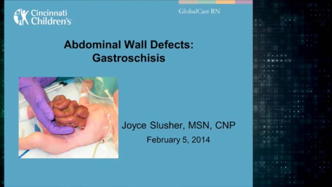
Gastroschisis: Advanced Practice Providers
43 min · Published Feb 2014
Only a few other public items share this expert — go deeper there →
Video
Pediatric Surgical Oncology Research Collaborative (PSORC): Studying Rare Pediatric Tumors
56 s · Published May 2026
Video
Update Course Rewind 2025: Hirschsprung + ARM: Rare but Real
1 min · Published May 2026
Video
Update Course Rewind 2025: Hirschsprung + ARM: Rare but Real
1 min · Published May 2026
Video
Pooling Patients to Study Rare Pediatric Tumors: An Introduction to PSORC
56 s · Published May 2026
Video
The fetal frontier: A review of current and emerging fetal therapies for genetic diseases
44 s · Published May 2026
Video
Indocyanine green assists with sentinel lymph node mapping in pediatric and adolescent patients
1 min · Published May 2026
What the experts said
Most umbilical hernias will close spontaneously in the first year and some in the second year.
Age 2 is the earliest Dr. Azarow would consider umbilical hernia repair, and only after extensive counseling; optimal timing is before school entry (age 4–5).
A long proboscis does not affect the decision to operate early on an umbilical hernia.
Larger umbilical defects (>1 cm) are less likely to close spontaneously, but this does not justify early surgery.
Race or ethnicity of the child does not affect the decision on timing of umbilical hernia repair.
True incarcerated umbilical hernias requiring emergency surgery must present with bowel obstruction symptoms (vomiting, inability to feed).
Red, tender umbilical lumps in children are usually incarcerated omentum, preperitoneal fat, or infected urachal cysts—not incarcerated bowel.
Incarcerated omentum or fat at the umbilicus can be treated with NSAIDs; infected urachal cysts with antibiotics.
Dr. Azarow cannot recall ever performing emergency surgery for an incarcerated umbilical hernia.
LMA anesthesia can be used for umbilical hernia repair if adequate abdominal wall relaxation is achieved, but this is anesthesiologist-dependent.
PDS or Maxon suture is preferred for umbilical hernia repair because it lasts twice as long as Vicryl and is non-braided (lower infection risk in semi-contaminated field).
Dr. Azarow uses prophylactic antibiotics for umbilical hernia repair but acknowledges this is anecdotal practice.
Umbilicoplasty (excision of redundant skin) should be performed during umbilical hernia repair to achieve a flat, cosmetically acceptable result.
Umbilicoplasty technique involves creating a pedicled skin flap with blood supply from above or below, avoiding complete circumferential incision.
Tacking the undersurface of umbilical skin to the fascia with braided suture (Vicryl) promotes inflammatory adhesion and prevents recurrent proboscis.
A pressure dressing should remain in place for 3 days after umbilical hernia repair.
Gastroschisis is actually an umbilical ring defect, not a paraumbilical defect, because the natural history is for the hole to close spontaneously (which only occurs at the umbilical ring).
Over half of gastroschisis patients closed with sutureless technique develop large umbilical hernias, but most close spontaneously over 2–3 years.
Suturing the gastroschisis defect makes the umbilical ring edge ischemic and destroys its integrity, leading to hernias that won't close spontaneously.
Omphalocele repair should be delayed as long as possible to allow abdominal domain expansion; operating too early is a mistake.
If mesh placement seems inevitable for omphalocele, wait longer—many can be primarily closed if given sufficient time.
Parents should watch for the child's flanks bulging when lying flat—this indicates the abdominal girth is expanding and primary closure may be achievable.
For large umbilical hernias in older teenagers, laparoscopic repair with mesh can be considered if primary closure seems unlikely.
Repair of small asymptomatic umbilical hernias is not absolutely indicated and can be watched indefinitely if the child has no symptoms.
A 5 mm umbilical defect very rarely causes intestinal incarceration; small pieces of fat may get caught and cause discomfort.
Infants with tiny drops of clear or whitish umbilical drainage should be watched for 6 months before considering surgery.
A Nebraska study showed that operating on umbilical drainage in the first 3 months increases complication rates; many resolve spontaneously by 6 months.
Dr. Azarow has stopped operating in the first 6 months for minor umbilical drainage or granulomas.
Umbilical granulomas can be treated with silver nitrate application; many resolve without surgery.
Office ligation of umbilical granulomas is not recommended unless the stalk is very narrow; they often don't fall off easily.
A prospective trial at Dr. Ponsky's institution found triamcinolone (Kenalog) cream superior to silver nitrate for umbilical granulomas, with such a drastic difference the study was stopped early.
Steroid cream works better than silver nitrate for granulation tissue at gastrostomy sites, so the same principle applies to umbilical granulomas.
For persistent umbilical drainage at 6–12 months, perform umbilical exploration through a hernia repair incision, divide the fascia inferiorly to visualize the urachal track, and bluntly dissect to the bladder dome.
The distal epithelialized portion of the urachal remnant must be excised from the umbilical skin, taking care not to devascularize the umbilicus.
A Nebraska study found no VCUG or ultrasound findings that changed management of urachal remnants, so Dr. Azarow does not perform preoperative imaging.
Infected urachal cysts can usually be treated with antibiotics alone; true abscesses require percutaneous drainage by interventional radiology.
After cooling down an infected urachal cyst, the entire congenital remnant should be excised electively to prevent recurrent abscess.
Elective urachal remnant excision is performed laparoscopically with a supraumbilical port and a 12 mm lateral port for stapling across the bladder dome.
Patent omphalomesenteric duct with stool draining from the umbilicus will not close spontaneously and requires surgery before hospital discharge.
Patent omphalomesenteric duct poses risk of volvulus from small bowel wrapping around the fistula tract.
Patent omphalomesenteric duct is approached through umbilical exploration; laparoscopy can assist if bowel is already twisted around the tract.
Epigastric hernias (epiploceles) are purely elective and should only be repaired if causing discomfort; they are preperitoneal fat through a pinhole defect, not true hernias.
The fascial defect in epigastric hernias is typically only 1 millimeter in diameter.
Analysis of PHIS (Pediatric Health Information System) data showed the mean age for umbilical hernia repair across U.S. children's hospitals is 4 years.
Emerging anesthesia data suggesting risks of operating before age 2–3 provides additional support for delaying umbilical hernia repair.
A Canadian study by Dr. Baird's group showed that gastroschisis patients closed with sutures had higher rates of subsequent umbilical hernia repair compared to sutureless closure.