
Hepatoblastoma
With Dr. Max Langham · hosted by Dr. Todd Ponsky · StayCurrentMD
Cued at 31:35 · stops at 32:20 · press play
Educational content from recorded physician discussions — not medical advice. Talk to your (or your child's) care team about your situation.
Video
Hepatoblastoma with Dr. Greg Tiao
CCHMC Pediatric Surgery · Published Mar 2022
Podcast
Hepatoblastoma with Dr. Greg Tiao
10 min · Published Feb 2022
Video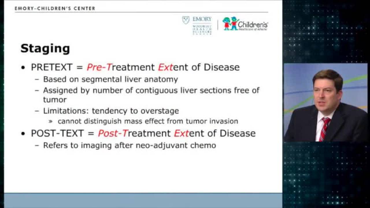
Hepatoblastoma: Update Course 2014
24 min · Published Nov 2018
Video
Social Determinants of Health Influence on Survival in Wilms Tumor, Neuroblastoma, and Hepatoblastoma
1 min · Published Jan 2026
Video
Quality of Life Outcomes for Patients Who Underwent Conventional Resection and Liver Transplantation for Locally Advanced Hepatoblastoma
1 min · Published Jan 2026
Podcast
Journal of Pediatric Surgery Article Review: June 2022, AAP Issue
15 min · Published Sep 2022
Only a few other public items share this expert — go deeper there →
Video
Pediatric Surgical Oncology Research Collaborative (PSORC): Studying Rare Pediatric Tumors
56 s · Published May 2026
Video
Update Course Rewind 2025: Hirschsprung + ARM: Rare but Real
1 min · Published May 2026
Video
Update Course Rewind 2025: Hirschsprung + ARM: Rare but Real
1 min · Published May 2026
Video
Pooling Patients to Study Rare Pediatric Tumors: An Introduction to PSORC
56 s · Published May 2026
Video
The fetal frontier: A review of current and emerging fetal therapies for genetic diseases
44 s · Published May 2026
Video
Indocyanine green assists with sentinel lymph node mapping in pediatric and adolescent patients
1 min · Published May 2026
What the experts said
Liver tumors are rare in children, and sorting out the diagnosis is crucial for good outcomes.
A one-year-old with a liver mass is likely to be hepatoblastoma, although rhabdoid tumor can occur at that age and is a bad actor.
If alpha-fetoprotein is quite high and very elevated, the presumptive diagnosis in a 1-year-old would be hepatoblastoma.
Hepatoblastoma presents from the first year of life through perhaps 10 years of age, although there are a few that have been diagnosed older than that.
If this is a rhabdoid tumor, chemotherapy is not important and the lesion needs to be resected.
If it's a very low risk hepatoblastoma, it probably doesn't need a biopsy and needs an upfront resection.
Most people favor needle biopsy of the liver rather than an open biopsy or a laparoscopic biopsy.
The biopsy should go through the normal part of the liver that will be resected with the specimen, approaching from the side of the tumor to avoid seeding the contralateral lobe.
Hepatoblastoma will seed and can create big issues for future resection and cure if the biopsy crosses the uninvolved lobe.
There have been prenatal diagnoses of hepatoblastoma, not super rare but not very common.
Rhabdoid tumor is more common in very young children compared to hepatoblastoma.
Hepatocellular carcinoma can occur in young children, usually in association with an inborn error of metabolism, with tyrosinemia as the prototype.
A baby with tyrosinemia will develop hepatocellular carcinoma if they are not transplanted, usually in the first couple of years of life.
There is a mixed tumor with features of hepatoblastoma and hepatocellular carcinoma that tends to occur in children over 6 years of age.
There is a predisposition of premature infants to getting hepatoblastomas, usually not in the nursery but in the first year or two of life.
For the biology arm of the Children's Oncology Group, they want 10 core biopsies.
The complication rate and some mortality seems higher with open biopsies than with the needle technique.
There is no role for frozen section in hepatoblastoma, either for initial biopsy or for later margin assessment.
The overarching goal of treatment for hepatoblastoma is to get the kids to a safe resection.
The PRETEXT system is a pre-treatment extent of disease radiographic imaging-based system developed in Europe during SIOPEL 1.
Couinaud based the numerology of liver segments on the street map of Paris, with the caudate lobe (segment 1) representing the Île de Paris where Notre Dame is located.
A PRETEXT 1 lesion has three contiguous sectors of the liver that are free of tumor.
A PRETEXT 2 lesion has two adjoining sectors free of tumor.
With PRETEXT 3, there is only one sector free of disease.
In PRETEXT 4, all of the sectors are involved with no sectors of the liver free of disease.
It is more common for people to over-read PRETEXT than under-read it.
In AHEP 0731 and the FIT trial, upfront resection is acceptable if it is a conventional hemihepatectomy or less, with clearly 1 centimeter of normal liver between the middle hepatic vein and tumor, and no annotation factors.
If a lesion is resected upfront and histology shows well-differentiated pure fetal histology, the baby is done with no need for any chemotherapy at all, and survival is 100%.
Along with imaging for suspected hepatoblastoma, a chest CT is needed because pulmonary metastases make up the vast majority of metastases, and even small liver tumors can metastasize.
If there is a PRETEXT 1 lesion with metastatic disease in the lungs, that is a high-risk patient.
For a PRETEXT 1 lesion with clear lungs, no vena cava, hepatic vein confluence, or portal vein involvement, and judged resectable by the surgeon, it should be resected with an anatomic resection and the child will be in a very low risk group.
A PRETEXT 1 lesion with vascular involvement (within 1 cm of cava or hepatic veins) would be considered an intermediate risk patient.
If a patient with hepatoblastoma is over 8 years of age, that moves the patient into a higher risk environment.
Not all PRETEXT 1 lesions are resectable upfront, and there are some PRETEXT 2 lesions that are resectable upfront.
Annotation factors include V for vena cava and hepatic veins; if they are more than 1 centimeter away from the vena cava, there is no annotation factor.
V0 means the lesion is within 1 centimeter of the cava or confluence of hepatic veins, making the patient no longer eligible for upfront resection.
V1 means the tumor is abutting either the vena cava or the three hepatic veins.
V2 means the tumor is large enough that it is distorting or pushing the vessels away.
V3 means there is evidence of tumor thrombus extending out of the vein, which is particularly problematic.
Hepatoblastoma is like Wilms tumor in that you can get direct extension of large tumor thrombus out of the hepatic veins into the vena cava and even up into the heart.
Most hepatoblastomas are a mixture of fetal and embryonal histologies.
The most common adverse finding is small cell undifferentiated (SCU) histology, which is a primitive tumor with small blue cell morphology and high nuclear-cytoplasmic ratio, making it a much higher risk lesion.
In AHEP 0731, patients with mixed fetal and embryonal histology after upfront resection with negative margins were treated with two courses of cisplatin-based chemotherapy with excellent results.
If the diagnosis is hepatoblastoma, the child needs long-term central access and will have two rounds of cisplatin-based chemotherapy in the United States, then be reimaged.
Most of the tumor shrinkage that will occur with chemotherapy happens in the first two rounds.
After two rounds of chemotherapy and reimaging, the surgeon should make a commitment about resectability; if resectable, do 2 more rounds of chemotherapy, resect after 4, then give 2 consolidation rounds for a total of 6.
Multifocal tumors should be referred to transplantation because they have high recurrence risk after resection.
Most surgeons will be able to make the call about resectability at the end of two rounds of chemotherapy.
A child that is 6 years of age or older with hepatoblastoma is going to be a significantly higher risk patient.
Children who are 8 years of age or older with hepatoblastoma are going to be high risk.
Children with hepatoblastoma who have normal or near-normal alpha-fetoproteins (less than 100) are going to be high-risk patients.
Pulmonary metastases in hepatoblastoma can have superb results, and current AHEP results look similar to Wilms tumor outcomes with pulmonary mets.
Rapid responders whose pulmonary mets disappear with chemotherapy probably do not need any specific pulmonary therapy.
A child where the pulmonary mets get smaller but persist will need a pulmonary metastectomy, which can be done thoracoscopically or open.
The Japanese have published using indocyanine green as a marker for thoracoscopic metastectomy because it is concentrated in the liver and lights up.
If pulmonary mets don't disappear, they should be biopsied to confirm metastatic disease and rule out other lesions like histoplasmosis in the Mississippi Valley.
Before doing anything of consequence with the liver, ensure you can vascularly isolate it by dissecting the suprahepatic and infrahepatic vena cava and placing umbilical tapes for potential clamping.
Disasters during liver resection happen because of major bleeding or air embolism from holes in the low-pressure vena cava system.
Use hypovolemic anesthesia with low CVP during liver resection, preserving transfusions until later in the case, because high CVP causes the liver to swell, become turgid, bleed more, and makes veins harder to dissect.
Take inflow before outflow during liver resection to prevent the liver from swelling.