
Educational content from recorded physician discussions — not medical advice. Always talk to your child's care team about your child's situation.
Content of this collection
Tools
0 items
High-Yield Summaries
1 item

Esophageal Atresia
Watch →
Comprehensive discussion of esophageal atresia and esophageal fistulas by Robert Parry. Initial management, operative management, pre operative echocardiogram, chest tube, thoracoscopic versus open esophageal atresia,TEF repair, long gap es
video39:30 · Nov 2018
In-depth Review
3 items


Tracheoesophageal Fistula with Dr. Daniel von Allmen
Listen →
This podcast discusses the work up, operative management, and follow-up management of tracheoesophageal fistulas. Dr. Todd Ponsky is an associate professor of surgery and pediatrics and pediatric surgeon at Akron Children's Hospital. Dr. Av
podcast45:21 · Dec 2020
Esophageal Atresia/Tracheoesophageal Fistula Bronchoscopy and Surgical Technique
Watch →
Is there any case that gets a pediatric surgery fellow more excited than the repair of an Esophageal Atresia with a Tracheoesophageal Fistula? In today's episode, we hear from Dr. Dan von Allmen, Dr. Mike Rutter, Dr. Aaron Garrison, and Dr.
video16:54 · Jul 2023
Treatment for Long Gap Esophageal Atresia: Esophageal Elongation and Replacement
Listen →
Pediatric surgeons Dr. Daniel Von Allmen and Dr. Todd Ponsky from Cincinnati children's Hospital discussing the technique for treating long gap esophageal atresia. For additional info please visit: https://www.youtube.com/c/CincinnatiChildr
podcast10:56 · Jan 2022
Work-up and Treatment
0 items
Technique Videos/OP Notes
2 items
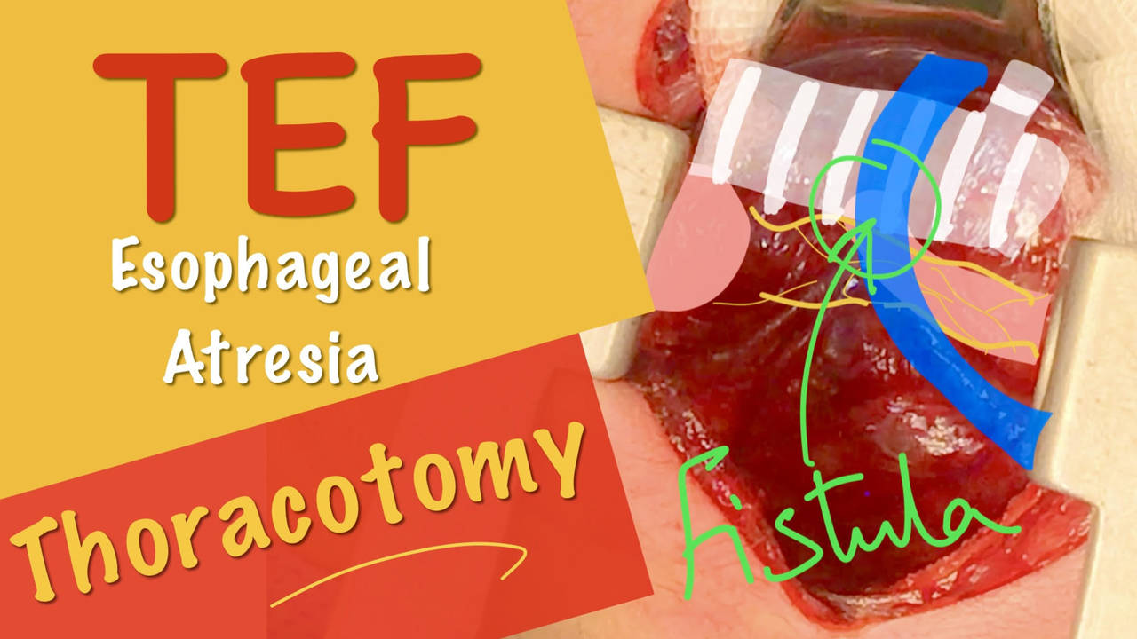

Neonatal Open Repair of Esophageal Atresia & Tracheo-esophageal Fistula EA_TEF. Dr Tamer Ashraf
Watch →
In this video, open thoracotomy and repair of esophageal atresia and distal tracheo-esophageal fistula is demonstrated in for a 3 day old baby. The video shows the surgeon's point of view (POV).
This video is intended as an education mater
video · Apr 2021
IPEG Academy: Thoracoscopic Esophageal Atresia Repair
Watch →
IPEG Academy Instructional Video: Thoracoscopic Esophageal Atresia Repair
M.C. Mora, D.A. Davies, H.D. Le, G. Azzie, K.A. Diefenbach
video · Jun 2021
Updates
2 items


Thoracoscopic Repair of Esophageal Atresia
Watch →
video · Oct 2023
Update Course Rewind: Magnet Therapy for Esophageal Atresia
Listen →
At Pediactric Surgery Update Course 2021, Dr. Bethany Slater from UChicago Comer Children's Hospital and Dr. Steven Rothenberg from Rocky Mountain Hospital for Children sharing their cases on using magnet therapy as an alternative treatment
podcast10:25 · Dec 2021
Summaries and takeaways+ Show
Summary of this collection+ Show
Key points, with the moment each was said+ Show
Featured picks for families are being prepared — the summaries above are written for you.
Takeaways+ Show
Esophageal atresia with tracheoesophageal fistula (TEF) remains a defining procedure in pediatric surgery, with challenges including long-gap atresia, recurrent fistulas, and strictures. Preoperative assessment centers on echocardiography to identify cardiac anomalies and aortic arch sidedness, though echo may misidentify arch anatomy; right-sided arch does not mandate left thoracotomy. Rigid bronchoscopy is increasingly standard, documenting fistula location, ruling out laryngeal cleft (easily missed and present in many referred patients with multiple failed repairs), and guiding endotracheal tube placement.[e300-c26–c28][e6895-c1–c4] The extrapleural right thoracotomy remains the workhorse approach, with azygos division providing access to the fistula. Thoracoscopic repair is feasible in ~75% of cases, offering superior visualization for proximal pouch mobilization but requiring advanced suturing skills and appropriate patient selection (>2 kg, uncomplicated cardiac disease).[e6895-c7–c9] Extensive proximal dissection—staying on the esophagus to avoid tracheal injury—yields the most length. Distal esophageal mobilization is safe laterally but risky medially.[e300-c42–c43] Fistula ligation must be flush with the trachea to prevent recanalization.[e6895-c17–c19] Transanastomotic tubes increase stricture and leak rates and are no longer recommended.[e300-c82–c83] For long-gap atresia (≥4 cm, gasless abdomen), G-tube placement with interval gap measurement guides timing of delayed primary repair; internal traction techniques (Foker, van der Zee) achieve 96% success in primary cases but require prolonged ICU stays and paralysis.[e4791-c7–c8] Magnet-assisted anastomosis (Flourish device) is FDA-approved for gaps <4 cm without fistula but carries high stricture rates.[e4763-c2–c6] Esophageal replacement (gastric pull-up or colonic interposition) is reserved for irreparable gaps.[e4791-c10–c12] Virtually all patients have reflux; fundoplication is indicated for recurrent strictures unresponsive to dilation.[e300-c105–c107] Long-term follow-up remains inadequate, with emerging data on Barrett's esophagus risk and swallowing dysfunction in adults.[e300-c97–c102]
- Rigid bronchoscopy is essential: it identifies laryngeal clefts (missed on flex scope), documents fistula location, and guides ET tube placement to optimize ventilation during repair.[e300-c26–c28][e6895-c1–c4]
- Thoracoscopic repair is feasible in 75% of cases and offers superior proximal pouch visualization, but requires advanced suturing skills; babies <2 kg and complex cardiac disease are relative contraindications.[e627-c13][e627-c16][e6895-c7–c9]
- Transanastomotic feeding tubes significantly increase stricture and leak rates and should be abandoned; this is supported by Midwest Pediatric Surgical Consortium data.[e300-c82–c83]
- For long-gap atresia, internal traction (Foker/van der Zee) achieves 96% primary success but requires weeks of paralysis and ICU care; magnet devices have high stricture rates and limited indications.[e4791-c7][e4763-c13]
- Virtually all EA patients have reflux; fundoplication is indicated for recurrent strictures. Long-term follow-up is inadequate—Barrett's esophagus risk and adult swallowing dysfunction require systematic study.[e300-c102–c107][e300-c97–c100]
For patients & families
Esophageal atresia (EA) is a birth defect where the esophagus — the tube that carries food from the mouth to the stomach — doesn't form properly. Most babies with EA also have a tracheoesophageal fistula (TEF), an abnormal connection between the esophagus and the windpipe. Doctors typically discover this condition shortly after birth when a feeding tube won't pass into the stomach, or sometimes before birth through ultrasound showing a small stomach and extra amniotic fluid. Before surgery, physicians perform an echocardiogram to check the heart and rule out other associated conditions that occur in many EA babies. The repair usually happens within the first few days of life through an operation that closes the abnormal connection and joins the two ends of the esophagus together. Surgeons can perform this repair through a small incision in the chest (thoracotomy) or using minimally invasive cameras and instruments (thoracoscopy). The medical team includes not just surgeons but also specialists in breathing, digestion, nutrition, and genetics who work together to care for these babies. Most children do well after repair, though some need additional procedures for complications like narrowing of the esophagus (stricture) or acid reflux.
Esophageal atresia (EA) is a birth defect where the esophagus — the tube that carries food from the mouth to the stomach — doesn't form properly. Most babies with EA also have a tracheoesophageal fistula (TEF), an abnormal connection between the esophagus and the windpipe. Doctors typically discover this condition shortly after birth when a feeding tube won't pass into the stomach, or sometimes before birth through ultrasound showing a small stomach and extra amniotic fluid. Before surgery, physicians perform an echocardiogram to check the heart and rule out other associated conditions that occur in many EA babies. The repair usually happens within the first few days of life through an operation that closes the abnormal connection and joins the two ends of the esophagus together. Surgeons can perform this repair through a small incision in the chest (thoracotomy) or using minimally invasive cameras and instruments (thoracoscopy). The medical team includes not just surgeons but also specialists in breathing, digestion, nutrition, and genetics who work together to care for these babies. Most children do well after repair, though some need additional procedures for complications like narrowing of the esophagus (stricture) or acid reflux.
The doctors in this collection+ Show
All expert statements+ Show
Every expert statement below comes from the recorded discussions, with its speaker and moment.
Treatment for Long Gap Esophageal Atresia: Esophageal Elongation and Replacement
At Cincinnati Children's, if a child has no gas in the abdomen, they place a G-tube and may put something up the distal esophagus for a fluoro shot, then wait a couple of weeks for a protocol gap measurement in interventional radiology.
clinicalDaniel von Allmen0:46 ↗
In the Foker technique, the surgeon ties sutures to either end of the esophagus through an open incision, brings them out through the chest wall, ties them together in a knot, and places spacers underneath periodically (about every day) until the ends come together for primary anastomosis.
Host summaryRod Gerardo summarizes what Dr. Daniel von Allmen said — not the host's own clinical position1:08 ↗
Stretch is a very strong promoter of growth, and if you put things on tension they will actually grow over time, which is how the cardiovascular system develops in utero.
clinicalDaniel von Allmen2:10 ↗
The philosophy of traction-based elongation is that with traction, you can get the two ends of the esophagus to grow, and if you can get them to grow far enough, you can put them together.
clinicalDaniel von Allmen2:10 ↗
The van der Zee technique uses the same concept as Foker but is done thoracoscopically with no external sutures, so all tension is inside the thorax.
Host summaryRod Gerardo summarizes what Dr. Daniel von Allmen said — not the host's own clinical position2:39 ↗
The Kimura technique involves creating a spit fistula and periodically moving it down the chest wall over time to stretch the proximal pouch closer to the distal pouch, but is not used all that often anymore.
Host summaryRod Gerardo summarizes what Dr. Daniel von Allmen said — not the host's own clinical position2:39 ↗
In the Boston Group 2015 study, the primary group (de novo cases) achieved an intact esophagus in 96% of patients, while the secondary group (patients with previous operations) achieved this in about two thirds of patients.
Host summaryDaniel von Allmen summarizing a resource — not the host's own clinical position3:15 ↗
In the Boston Group 2015 study, the primary group had a median ICU stay of 70 days with a couple of weeks being paralyzed, and the secondary group had a median ICU stay of 110 days with a month of being paralyzed.
Host summaryDaniel von Allmen summarizing a resource — not the host's own clinical position3:15 ↗
In the Boston Group 2015 study, about two thirds of patients with primary repair were able to get full oral nutrition and about 10% of patients who had secondary repair achieved full oral nutrition.
Host summaryDaniel von Allmen summarizing a resource — not the host's own clinical position3:15 ↗
The surgical group from INOEA recommends gastric pull up as the first option for esophageal replacement, dividing the esophageal stump at the esophageal hiatus and mobilizing the fundus to pull it up in either the anterior or posterior mediastinum.
Host summaryDaniel von Allmen summarizing a resource — not the host's own clinical position4:36 ↗
The colon can be used as an interposition for esophageal replacement.
clinicalDaniel von Allmen4:36 ↗
In colonic interposition, the piece of colon chosen is based on the blood supply and the diameter needed.
Host summaryRod Gerardo summarizes what Dr. Daniel von Allmen said — not the host's own clinical position5:08 ↗
In colonic interposition, a pyloroplasty is performed to help with gastric emptying.
Host summaryRod Gerardo summarizes what Dr. Daniel von Allmen said — not the host's own clinical position5:08 ↗
Common problems with colonic interposition are that the colon can dilate and become tortuous, and it's not uncommon to get a sigmoid sink drain deformity just above the diaphragm.
clinicalDaniel von Allmen8:00 ↗
Von Allman was initially taught that sigmoid redundancy in colonic interposition can't be fixed and that it's too dangerous because it will risk the blood supply, but he found that this is not really true.
opinionDaniel von Allmen8:17 ↗
Von Allman passes the colonic interposition posterior to the stomach, which leaves the vascular pedicle along the spine, allowing mobilization of the colon by dividing the gastric duodenotomy and colon attachment to the stomach, then mobilizing the sigmoid redundancy transhiatally and reanastomosing the colon to the stomach.
clinicalDaniel von Allmen8:17 ↗
Colonic interpositions sometimes need to be revised, but it is not impossible to revise them and the kids tend to do pretty well.
opinionDaniel von Allmen8:17 ↗
You can get extraordinary length with colonic interposition, allowing treatment of cases with caustic injuries extending to the pharynx by sewing the colon to the pharynx and down to the stomach, which is tough to do with a gastric pull-up.
clinicalDaniel von Allmen9:03 ↗
The short interval between time of traction and anastomosis (less than five days) raises the question of whether the mechanism is stretching or growth, which will impact results and likely reflects on complications.
Host summaryRod Gerardo summarizes what Dr. Daniel von Allmen said — not the host's own clinical position9:27 ↗
Von Allman states we don't know whether esophageal elongation is growth or stretch and should do studies to understand that, but notes that tension is a very good physiologic growth promoter in other organs, making this an area ripe for more basic science.
opinionDaniel von Allmen9:56 ↗
Tracheoesophageal Fistula with Dr. Daniel von Allmen
TEF is a defining procedure of pediatric surgery with challenges including long gap atresia, recurrent fistulas, and strictures
Host summaryThe host summarizes what Dr. Daniel von Allmen said — not the host's own clinical position0:00 ↗
Cincinnati Children's has a multidisciplinary aerodigestive center including ENT airway surgeons, GI, pulmonary, general surgeons, nurse practitioners, geneticists, and dietitians
clinicalDaniel von Allmen2:01 ↗
The aerodigestive center typically receives more complicated patients referred from outside through either esophageal or airway arms
clinicalDaniel von Allmen2:46 ↗
Physical examination should assess for scaphoid versus full or distended abdomen and respiratory distress
clinicalDaniel von Allmen3:55 ↗
The surgeon should personally attempt to pass the NG tube rather than relying on nursing reports, as tubes reported as not passing sometimes pass easily
clinicalDaniel von Allmen4:20 ↗
Injecting air into the NG tube and obtaining a plain film helps visualize proximal pouch distention and assess distal GI gas pattern
clinicalDaniel von Allmen4:55 ↗
Lack of visible distended proximal pouch raises concern for either incorrect diagnosis or presence of proximal fistula allowing pouch decompression
clinicalDaniel von Allmen5:01 ↗
Contrast studies can be performed if there is high suspicion of unusual anatomy, as long as the radiologist knows the esophagus may be obstructed
clinicalDaniel von Allmen5:42 ↗
Echocardiogram is the most important preoperative study to assess cardiac anatomy and rule out right-sided aortic arch
clinicalDaniel von Allmen6:25 ↗
Renal ultrasound and spine evaluation for tethered cord are needed but are elective and not necessary before addressing esophageal atresia
clinicalDaniel von Allmen6:54 ↗
What's new+ Show