


From
StayCurrentMD
Multi-modal 3-Dimensional Visualization of Pediatric Neuroblastoma: Aiding Surgical Planning Beyond Anatomical Information
Part of
Pediatric Oncology 696 items
jpedsurg.org shows its articles on its own site.
Read the article on jpedsurg.org ↗Article · Feb 2024 · 1 min read
In brief
In brief
This work presents a novel workflow combining anatomical 3D modeling with functional imaging (ADC and MIBG-SPECT-CT) to create preoperative heatmaps that distinguish viable from non-viable neuroblastoma tissue. The multimodal approach addresses the limitation of traditional anatomical models by incorporating tumor biology, potentially improving surgical decision-making in pediatric neuroblastoma resection.
Written by the GCMD Library team from the article.
Patient-specific 3D models of neuroblastoma and relevant anatomy are useful tools for surgical planning. However, these models do not represent the heterogenous biology of neuroblastoma. This heterogeneity is visualized with the ADC and 123I-MIGB-SPECT-CT imaging. Combining these multi-modal data into preoperative 3D heatmaps, may allow differentiation of the areas of vital and non-vital tumor tissue. We developed a workflow to create multi-modal preoperative 3D models for neuroblastoma surgery.
Educational content from recorded physician discussions — not medical advice. Talk to your (or your child's) care team about your situation.
Video
Oncology Tumor Board Showdown: Pediatric Oncology 5 Question Quiz
1 min · Published Oct 2026
Article
Minimally invasive surgery for abdominal and thoracic neuroblastic tumors: A systematic review by the APSA Cancer committee
Juan Pablo Gurria · Published Oct 2026
Video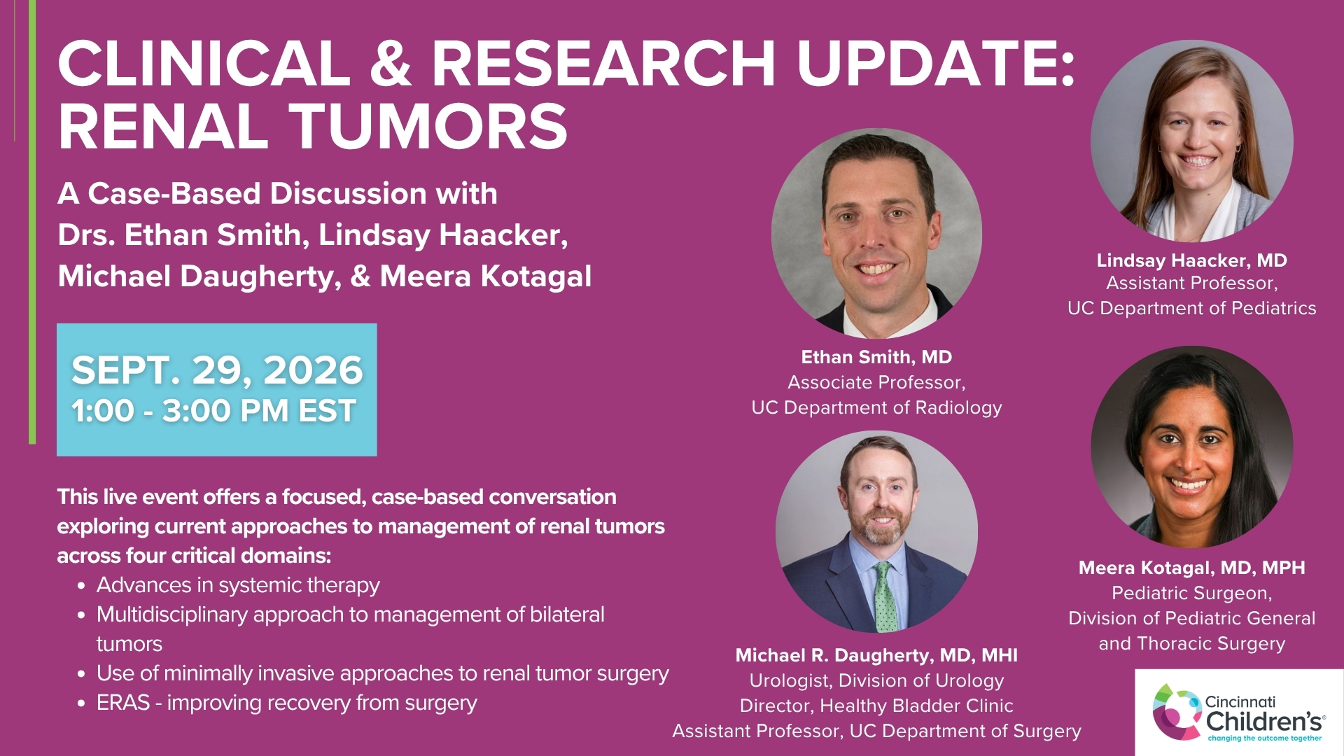
Clinical & Research Update: Renal Tumors with Drs. Ethan Smith, Lindsay Haacker, Michael Daugherty, and Meera Kotagal
71 min · Published Sep 2026
Video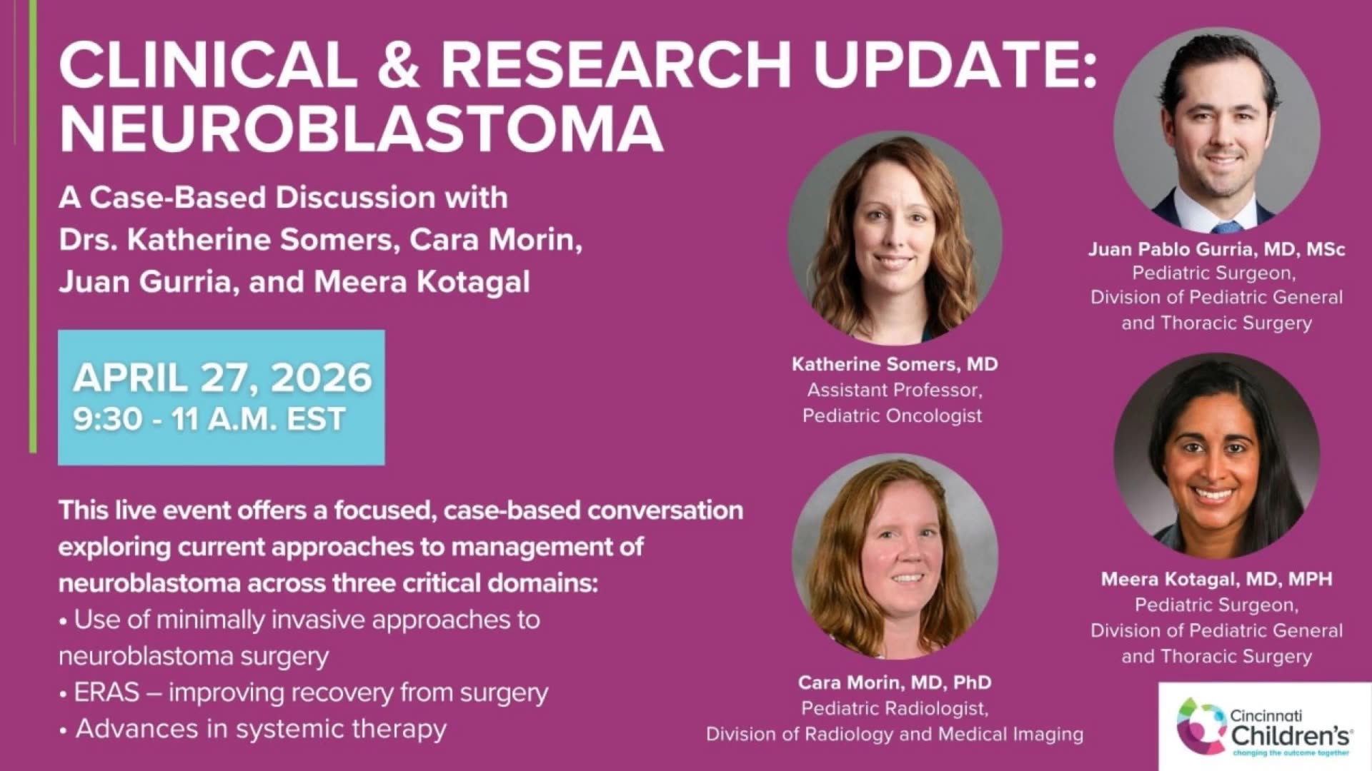
Clinical & Research Update: Neuroblastoma with Drs. Katherine Somers, Cara Morin, Juan Gurria, and Meera Kotagal
67 min · Published Sep 2026
Video
Clinical & Research Update: Sarcoma w/ Drs. Roshni Dasgupta, Joseph Pressey, Arthur Meyer, Luke Pater
66 min · Published Sep 2026
Video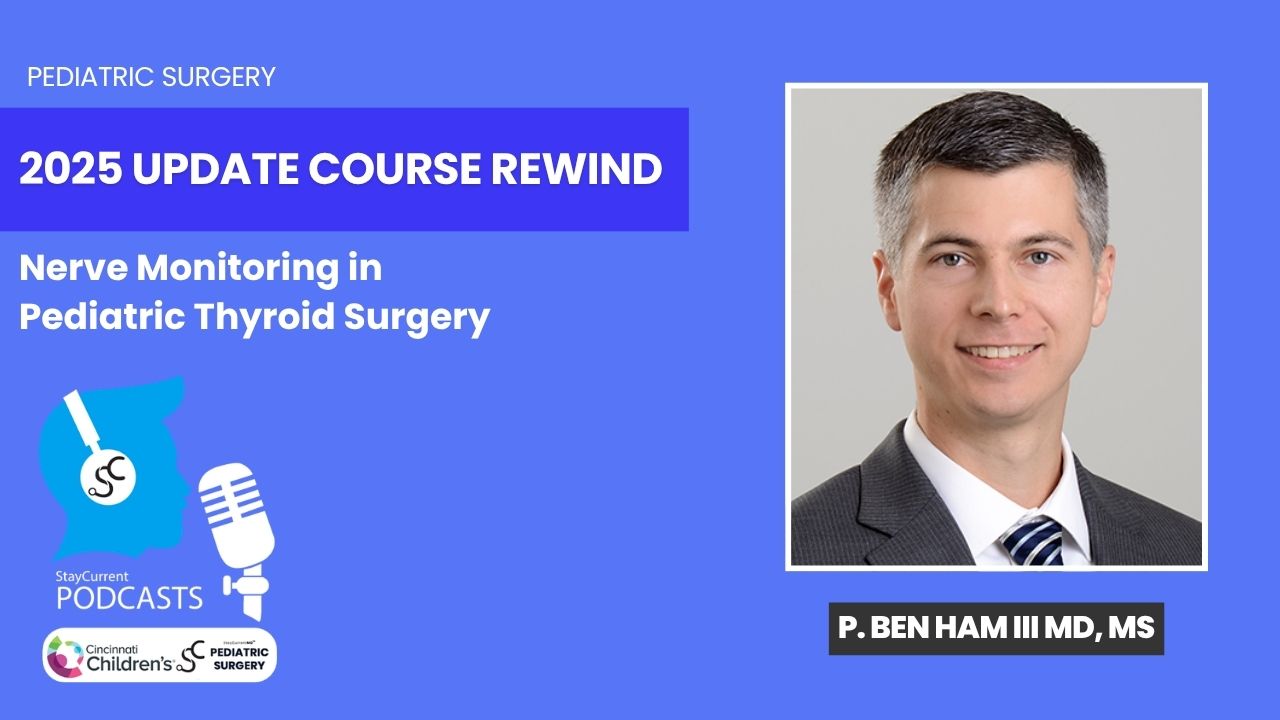
Update Course Rewind 2025: Nerve Monitoring in Pediatric Thyroid Surgery
6 min · Published Jul 2026
Video
Intestinal Atresia Types Explained: Grossfeld Classification for Pediatric Surgery
1 min · Published Sep 2026
Video
Derivation and validation of the Pediatric Community-Acquired Pneumonia Severity (PedCAPS) score: A prospective cohort study
50 s · Published Sep 2026
Video
FETO for Late-Diagnosed Severe Congenital Diaphragmatic Hernia (CDH) at Cincinnati Children's with Dr. Beth Rymeski
4 min · Published Sep 2026
Video
Severe Congenital Diaphragmatic Hernia (CDH) Case: FETO Management & Unexpected Findings with Dr. Beth Rymeski
3 min · Published Sep 2026
Video
Fetoscopic Endoluminal Tracheal Occlusion (FETO) Procedure: Step-by-Step Guide with Dr. Beth Rymeski
4 min · Published Sep 2026
Video
Choledochal Cyst Types Explained: Pediatric Surgery Fundamentals
1 min · Published Sep 2026