

Wilms Tumor
With Dr. Peter Ehrlich & Dr. Abdul Raoof Lamoshi · hosted by Dr. Em Gootee & Dr. Todd Ponsky
Part of
Nephroblastoma 3 items
Chapter 3 of 14 · Diagnosis & Workup
Workup strategy
Initial Workup and Indications for Primary Surgery vs Chemotherapy
Cued at 6:23 · press play
Expert statements on this page
No expert statements were drawn from this page.
Host summaries · secondary, not cited in answers
Educational content from recorded physician discussions — not medical advice. Talk to your (or your child's) care team about your situation.
Podcast
Topics in 10: Wilms Tumor
12 min · Published Aug 2019
Video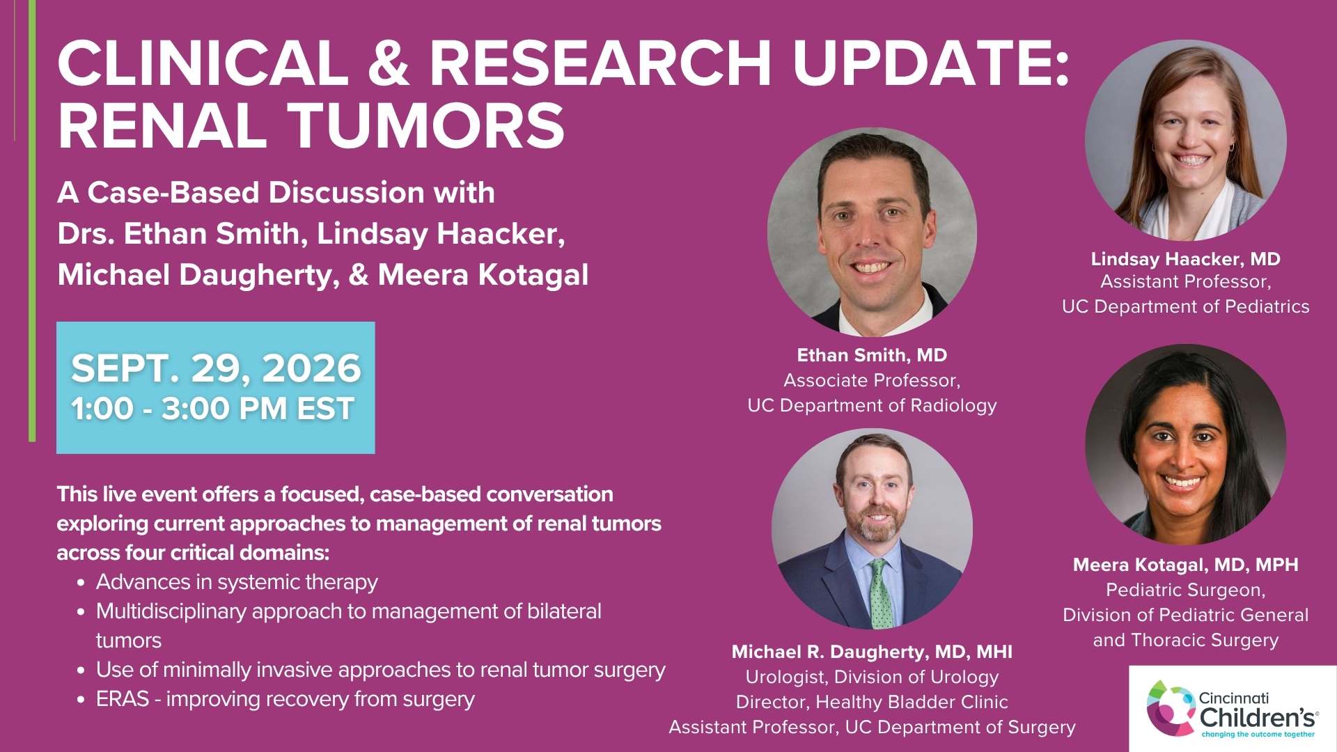
Clinical & Research Update: Renal Tumors with Drs. Ethan Smith, Lindsay Haacker, Michael Daugherty, and Meera Kotagal
71 min · Published Sep 2026
Video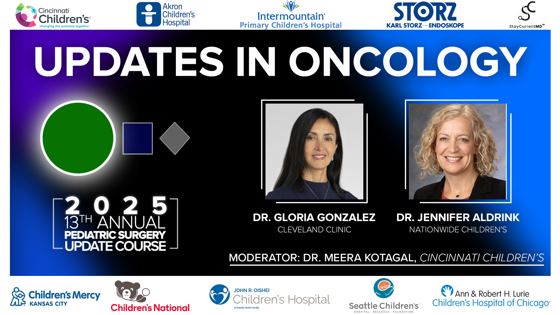
2025 Pediatric Surgery Update Course - Updates in Oncology
21 min · Published Aug 2025
Video
Wilms Tumor Protocol Violations: Practice Gap discussion at Update Course 2018
9 min · Published Aug 2018
Podcast
Neuroblastoma
56 min · Published Aug 2019
Video
Update Course Rewind: Updates in Wilms Management 2024
4 min · Published Sep 2025
What the experts said
In North America, the Children's Oncology Group recommends primary nephrectomy and ureterectomy with lymph node sampling for the majority of renal tumors in children.
Preoperative chemotherapy is recommended when tumor extends into the IVC beyond the infrahepatic level, as complication rates are higher with primary surgery in these cases.
Preoperative chemotherapy is indicated if the tumor is so large that major liver or bowel resection would be required, because the majority will respond to chemotherapy and complication rates are higher when these organs must be resected.
Children with bilateral Wilms tumors, solitary functioning kidney, or genetic predisposition syndromes (Wagr, Denys-Drash, Beckwith-Wiedemann) should receive preoperative chemotherapy rather than primary nephrectomy.
In situations requiring preoperative chemotherapy, biopsy should be performed to determine histology, as chemotherapy differs for favorable vs unfavorable histology and for different tumor types (Wilms, rhabdoid, clear cell sarcoma).
For biopsy, either open biopsy or large core sampling with at least 10 cores is recommended; accuracy increases between 10 and 20 cores. Fine needle aspiration or simple true-cut biopsy is insufficient because anaplasia cannot be diagnosed from these samples.
A central venous catheter or port should be placed at the time of biopsy to avoid multiple anesthetics; a single lumen is preferred due to lower infection risk.
COG staging distinguishes abdominal stage from disease stage. Stage 1 is tumor limited to kidney, completely resected, no capsular invasion, no rupture or biopsy, vessels not involved, negative margins and nodes.
Stage 2 is complete resection with tumor extending beyond kidney through capsular penetration, renal sinus invasion, or blood vessel involvement within the nephrectomy specimen outside the primary kidney.
Stage 3 includes: biopsy with gross residual tumor, positive lymph nodes, peritoneal surface penetration with implants, positive margins, microscopic residual from spillage, incomplete resection, piecemeal removal, or dividing the renal vein with tumor present.
Stage 4 is hematogenous metastasis to lung, liver, bone, or brain. Stage 5 is bilateral renal involvement.
A patient can have stage 1 or 2 abdominal disease with lung metastases (stage 4 disease stage). Such patients receive chemotherapy for metastatic disease but do not require abdominal radiation if abdominal stage is 1 or 2.
Stage 1 and 2 abdominal disease without metastases receives two-drug chemotherapy (vincristine and dactinomycin) for 19 weeks, which has lower toxicity and significantly lower risk of late effects than three-drug therapy with doxorubicin or radiation.
The main late effects of Wilms tumor treatment are renal failure, second malignancies, pregnancy complications in females, hypertension, and cardiovascular disease. The two main contributing factors are radiation and doxorubicin.
If a patient has a lung lesion but the abdominal tumor is resectable, primary nephrectomy should still be performed. Biopsying the abdominal tumor would automatically create stage 3 disease requiring abdominal radiation, whereas removing it with negative nodes avoids abdominal radiation.
A recent COG trial showed that 40% of patients with pulmonary metastases who achieve complete response by 6 weeks of chemotherapy do not need pulmonary radiation, with 80-85% remaining disease-free. Those who relapse can be salvaged with radiation.
Fifteen percent of girls who receive pulmonary radiation for Wilms tumor develop breast cancer, making radiation avoidance in responders a significant benefit.
For patients with lung lesions and resectable abdominal tumor, the approach is to perform nephrectomy and start chemotherapy based on kidney pathology without biopsying the lung lesion initially. At 6 weeks, if a single lesion remains or lesions don't respond, thoracoscopic biopsy may be performed, as 50-60% of residual lesions are not cancer.
All patients with lung lesions presumed to be metastatic disease start with three-drug chemotherapy.
The SIOP protocols used in Western Europe give preoperative chemotherapy to all patients without biopsy, using higher doses of vincristine and dactinomycin than COG protocols, with response evaluation at 4 and 8 weeks before proceeding to nephrectomy.
SIOP uses post-chemotherapy, post-nephrectomy classification into low, intermediate, and high risk. High-risk patients receive abdominal radiation regardless of lymph node status; intermediate-risk patients receive radiation if lymph node positive.
In SIOP protocols, predominantly blastemal component after preoperative chemotherapy is a negative prognostic factor, but blastemal percentage does not correlate with outcome in COG primary nephrectomy patients.
There is no outcome difference between patients who undergo empiric biopsy versus those who have intraoperative spillage—both are stage 3. The main prognostic factor in stage 3 is lymph node status.
Outcomes for stage 1 and 2 patients are essentially identical between COG and SIOP approaches.
Preoperative assessment should include hemoglobin (some tumors rupture internally causing anemia), PT/PTT (acquired von Willebrand disease can occur, rarely causing significant bleeding), and electrolytes.
Surgical approach uses transverse or modified subcostal incision, not median or retroperitoneal incisions. The tumor is mobilized completely before identifying vessels, as attempting early hilar dissection on large tumors can be dangerous.
Lymph node sampling should aim for at least 5-6 nodes from the renal hilum and para-aortic/paracaval regions, though evidence for a specific number is limited.
Taking the renal artery before the vein is preferred by principle but not always possible with large tumors. The order of vessel division does not significantly impact outcomes.
Positive surgical margins automatically make the tumor stage 3. Microscopic rupture through the capsule that is only visible on pathology also creates stage 3 disease.
Taking portions of adherent diaphragm or liver to avoid tumor violation does not upstage the tumor if true invasion has not occurred—these tumors often cause intense inflammatory reaction that mimics invasion.
Very low-risk patients (stage 1, favorable histology, tumor <550g, age <2 years) can be treated with surgery alone. This group has 90-95% cure rate without chemotherapy, and the 5-10% who relapse have 100% salvage rate.
IVC tumor extension is not a negative prognostic factor if properly managed. Tumors extending into the renal vein that come out in one piece are considered stage 2.
For IVC thrombus below the infrahepatic level, intraoperative ultrasound should be used to assess extent. The ideal technique is to mobilize the kidney, ligate the renal artery and accessory veins, make a small nick in the renal vein, and slide the thrombus out intact.
Tumors extending above the infrahepatic IVC have significantly higher complication rates (26-30% major morbidity) with primary surgery and should receive preoperative chemotherapy.
After reviewing 6000 operative notes, cases of tumor thrombus completely filling the IVC but stopping at the infrahepatic level are extremely rare—when the IVC is truly filled, the thrombus typically extends higher.
Favorable histology Wilms tumor has three components: blastemal, stromal, and epithelial (triphasic). Tumors with only two components are also considered favorable. In COG protocols, the amount of blastema has never correlated with outcome.
Stage 1 and 2 favorable histology without metastases receives two-drug chemotherapy (vincristine and dactinomycin) for 19 weeks. Stage 3 adds doxorubicin (DD4A regimen) for 25 weeks.
Loss of heterozygosity (LOH) testing is performed on all favorable histology tumors. Patients with both 1p and 16q LOH (5-7% of cases) have significantly worse outcomes: 10% lower survival for stage 1-2, 18% lower for stage 3-4.
Patients with both 1p and 16q LOH receive intensified treatment: stage 1-2 patients add doxorubicin (three-drug therapy), and stage 3-4 patients receive regimen M (five-drug chemotherapy).
Unfavorable histology is classified as focal anaplasia or diffuse anaplasia based on the number of high-power fields showing anaplasia. Treatment ranges from three-drug to five-drug chemotherapy plus or minus radiation depending on extent.
Other renal tumors include renal cell carcinoma (second most common, no good therapy for metastatic disease), clear cell sarcoma (reasonable treatment for low stages), and rhabdoid tumor (poor outcomes except stage 1; most present stage 3-4 with terrible prognosis).
Bilateral Wilms tumors occur in 8-10% of cases. The strategy is preoperative chemotherapy to shrink tumors enough to allow at least one partial nephrectomy, avoiding dialysis.
Historical outcomes for bilateral Wilms were significantly worse than unilateral: 61% event-free survival and 80% overall survival for bilateral vs 88% event-free and 95% overall for unilateral.
The recent COG bilateral Wilms study used VAD induction chemotherapy with imaging at 6 and 12 weeks. Maximum tumor response typically occurs by 12 weeks, and early response at 6 weeks predicts late response.
In typical-age children (<36 months) with bilateral renal tumors, biopsy is not mandatory as it is almost universally Wilms tumor. Of 250 patients enrolled in the recent study, only one had rhabdoid tumor (and the smaller lesion responded to therapy).
Biopsy is recommended for bilateral tumors in older children (8-10 years) or those with syndromes like von Hippel-Lindau where renal cell carcinoma is more likely. If biopsy is performed, both kidneys should be biopsied as there is discordant pathology in up to 20% of cases.