


From
Live Event Content
Update Course 2023 - Updates in the use of ICG
With Dr. Seth Goldstein · hosted by Dr. Tom Isch
Chapter 2 of 6 · Surgical Management
Depth & laparoscopy
Technical Considerations and Laparoscopic Applications
Cued at 8:40 · press play
Expert statements on this page
No expert statements were drawn from this page.
Host summaries · secondary, not cited in answers
Educational content from recorded physician discussions — not medical advice. Talk to your (or your child's) care team about your situation.
More about this diagnosis
Alveolar rhabdomyosarcomaVideo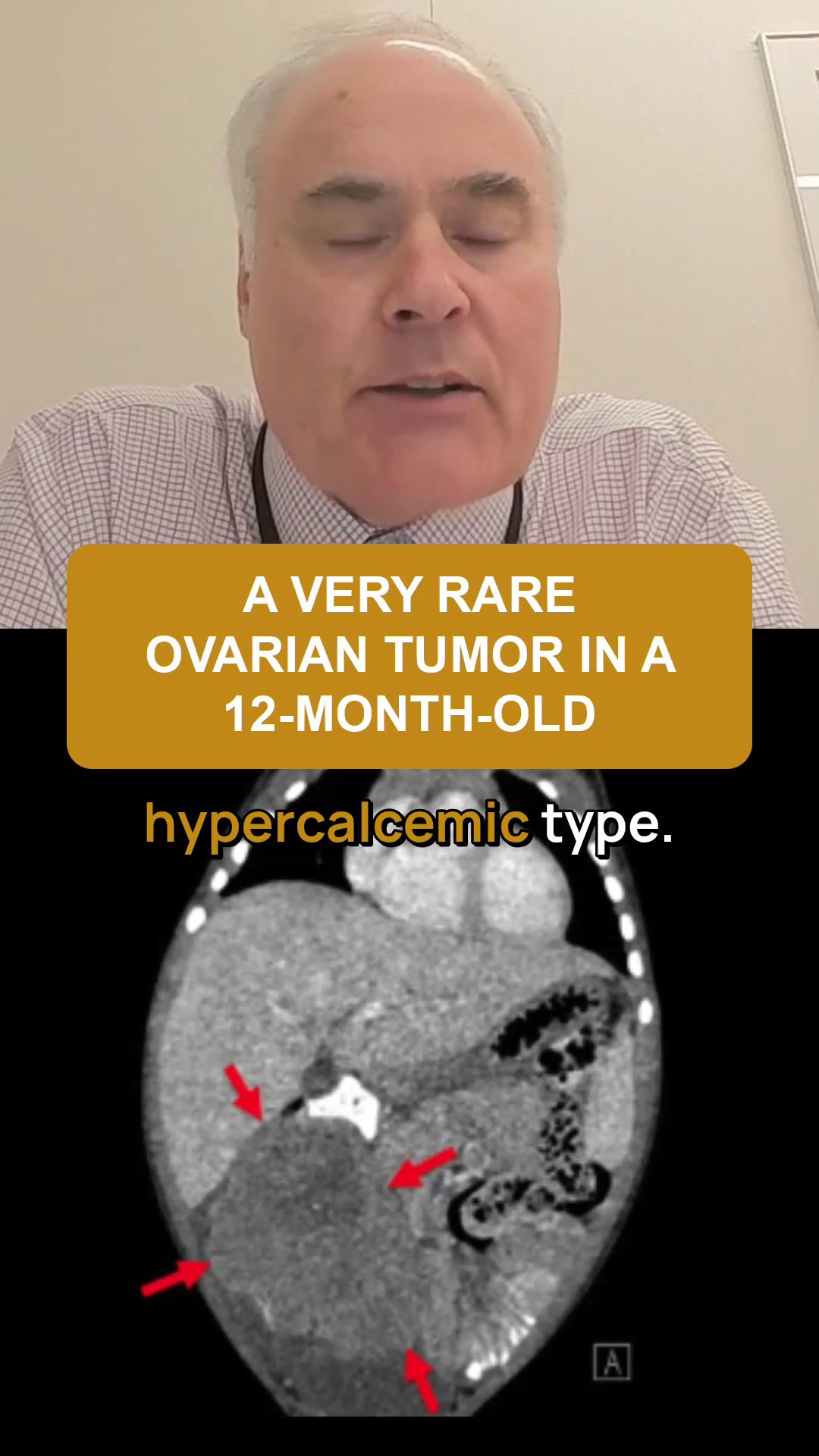
Rare Ovarian Tumor in Infants: Small Cell Carcinoma of Ovary, Hypercalcemic Type & SMARCA4 Mutation
21 s · Published Oct 2026
Video
Can Normal X-Rays Miss Metastatic Neuroblastoma? A Pediatric Radiology Case Study
26 s · Published Oct 2026
Video
Can Fractures Be a Sign of Hepatoblastoma in Children? Understanding Pediatric Liver Cancer Symptoms
37 s · Published Oct 2026
Video
Pancreas, Muscle, and Subcutaneous Fat Atrophy in Patients Undergoing Radiation for Neuroblastoma
47 s · Published Oct 2026
Podcast
Hepatoblastoma
Todd Ponsky · 54 min · Published Oct 2026
Podcast
Wilms Tumor
Todd Ponsky · 64 min · Published Oct 2026
Video
Clinical & Research Update: Renal Tumors with Drs. Ethan Smith, Lindsay Haacker, Michael Daugherty, and Meera Kotagal
71 min · Published Sep 2026
Video
Clinical & Research Update: Neuroblastoma with Drs. Katherine Somers, Cara Morin, Juan Gurria, and Meera Kotagal
67 min · Published Sep 2026
Video
Clinical & Research Update: Sarcoma w/ Drs. Roshni Dasgupta, Joseph Pressey, Arthur Meyer, Luke Pater
66 min · Published Sep 2026
Video
2026 Laparoscopic Pediatric Hernia Repair
123 min · Published Jun 2026
Video
Beyond the Spectrum: Diagnosis, Myths & Management - Caitlin Couch & Leslie Lopez - APP Conference 2026
50 min · Published May 2026
Video
Dysautonomia: Navigating the Journey - Martha Willis - APP Conference 2026
55 min · Published May 2026
What the experts said
Sentinel node biopsy in pediatric rhabdomyosarcoma is standard of care and dictates drug regimen and indicates radiation to lymph node basin if positive
ICG can be used as an adjunct to sentinel node identification in pediatric surgical oncology
ICG technique allows real-time intraoperative control of injection location (intratumoral vs. four quadrants, dermal vs. subdermal) by surgeons rather than radiologists
ICG fluorescence visualization occurs over 45-75 seconds after injection, allowing direct observation of lymphatic drainage to sentinel node
Near-infrared fluorescence penetrates approximately 1-2 centimeters through soft tissue
ICG enables laparoscopic sentinel node identification in paratesticular rhabdomyosarcoma, which is not feasible with radiotracer methods
In children under 10 years old with paratesticular rhabdomyosarcoma, sentinel node biopsy may be performed instead of full ipsilateral lymph node clearance
Indocyanine green was developed by Kodak company after World War II and has been FDA approved since the 1960s
ICG is approved for all adult and pediatric applications with no on-label or off-label concerns
ICG is exclusively cleared through the biliary system
For hepatic metastases in the chest, ICG administered 24 hours preoperatively will be retained only in liver tissue by the time of thoracotomy, enabling metastasis localization
ICG technique for hepatic metastases is successful for hepatoblastoma but has not been successful for Wilms tumor
Sarcomas other than hepatic primaries require much higher ICG dosage and are less specific
Wilms tumor metastases in the lungs are avid for ICG, though the primary kidney tumor is not
ICG fluorescence identifies additional pulmonary metastases beyond what visual inspection and palpation detect during thoracotomy
Infrared ureteral stents emit light that travels through centimeters of tissue and can be detected by standard fluorescence imaging systems
A renally-cleared fluorophore is under investigation that could enable ureteral identification without stent placement
ICG is used for vascular mapping in partial nephrectomy for Wilms tumor
ICG can identify accessory cystic ducts during cholecystectomy by showing separate fluorescence of the accessory duct, main cystic duct, and common bile duct