12 views 0 likes

StayCurrentMD

GCMD Space · View profile →

Tricks - Imperforate Anus and Rectourethral Fistula

Video Published 2018-11-10 Updated 2026-08-01

Timestops (4)

Topic Overview

A technical discussion of laparoscopic repair techniques for anorectal malformations with rectourethral fistulas and thoracoscopic tracheoesophageal fistula repair. The presenter describes a novel intraoperative measurement technique using calibrated catheters and simultaneous cystoscopy/colonoscopy to determine residual fistula length, aiming to achieve complete excision (≤5mm residual) while avoiding injury to surrounding structures. For TEF repair, a technique of leaving partial attachments of both proximal and distal esophageal ends during anastomosis is presented to facilitate handling and prevent retraction. Discussion includes trocar positioning strategies, bladder decompression methods, colostomy placement preferences, and clip application techniques.

Key Takeaways

  • Intraoperative catheter measurement ensures residual rectourethral fistula ≤5mm, preventing diverticula (0/23 cases at 2yr f/u). (2:37)
  • Bulbar fistulas require tube vesicostomy for bladder decompression and closer trocar placement vs prostatic fistulas. (6:16)
  • Sigmoid colostomy at descending junction provides adequate length for pull-through; transverse has infection/prolapse risks. (17:19)
  • For TEF repair, leaving 25% of distal fistula and 12-15% of proximal tip uncut prevents retraction during anastomosis. (28:00)
  • Clips applied too tightly crush muscle and cause fistula recurrence; transfixing suture closure is preferred. (37:16)

Inside this episode

Kai, the Library's AI content creator, listened to this episode and mapped who's speaking, the chapters, key claims, and cases. Every item links to the exact moment in the recording.

AI-enriched

Who's speaking

  • Speaker 1 — host
  • Speaker 2 — guest
  • Jeff Blair — guest
  • Speaker 4 — guest
  • Speaker 5

Chapters

  • 0:00Laparoscopic Repair of Imperforate Anus with Rectourethral Fistula — Introduction of novel fistula length measurement technique using calibrated catheters, colonoscopy, and cystoscopy. Demonstration of prostatic fistula case with trocar positioning and dissection approach. Emphasis on achieving residual fistula length ≤5mm through iterative measurement and mucosectomy.
  • 10:51Discussion: Laparoscopic ARM Repair Techniques — Faculty discussion of colostomy placement (transverse vs sigmoid), trocar positioning, bladder decompression methods, learning curve considerations, and comparison with PSARP approach. Debate over applicability to bulbar fistulas and technical complexity for general pediatric surgeons.
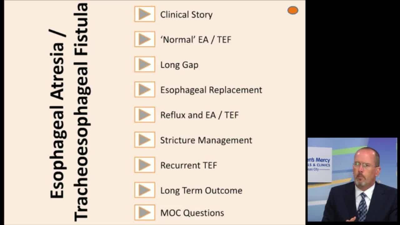
  • 25:52Thoracoscopic TEF Repair Technique — Presentation of technique leaving partial attachments of fistula and proximal esophageal tip during anastomosis to prevent retraction and facilitate handling. Demonstration of gap measurement using bronchoscopy, anastomotic technique grabbing uncut portions, and sequential division after initial sutures placed.
  • 35:38Discussion: TEF Repair Approaches and Clip Use — Faculty discussion of thoracoscopic vs open TEF repair, clip application techniques and erosion concerns, alternative anastomotic methods including transfixing sutures and sliding knots, and handling of esophageal tissue during dissection.

Key claims

  • 0:21Dissection of the fistula up to the red line is necessary to prevent residual fistula without injuring nerves, prostate, urethra, and sphincters — Speaker 1
  • 0:5123 male patients with rectourethral fistula were studied: 1 vesical, 14 prostatic, 9 bulbar, 5 no fistula — Speaker 1
  • 1:52A fine flexible colonoscope inserted into anterior rectal wall allows observation of both fistula orifice and level of laparoscopic dissection intraluminally — Speaker 1
  • 2:37A calibrated catheter inserted through the fistula opening while another surgeon performs cystoscopy allows measurement of inside fistula length between rectal opening and urethral orifice — Speaker 1
  • 4:27If residual fistula length is longer than 5mm, the rectal end is further dissected toward urethra using mucosectomy to prevent injury of prostate and urethra — Speaker 1
  • 4:57The measurement and dissection procedure is repeated until residual fistula length is ≤5mm, then fistula is ligated and excised — Speaker 1
  • 6:16For bulbar fistula, tube vesicostomy to decompress bladder is very important to obtain clear surgical field of deep pelvic floor — Speaker 1
  • 6:49For bulbar fistula, right and left trocars are placed much closer to telescope compared to prostatic fistula, which is key for reaching deep pelvic structures — Speaker 1
  • 8:30After fistula is tied, catheter is reinserted to gently probe tied fistula, allowing surgeon to reconfirm residual fistula length is ≤5mm — Speaker 1
  • 9:25In first 8 cases, initial fistula measurements from rectal to urethral orifice ranged from 5-21mm; 7 cases required further dissection, 1 did not — Speaker 1
  • 9:48During cystoscopy, normal saline refluxed into pelvic floor through fistula in 6 cases (indicating large fistula), but no reflux in 2 cases (indicating very narrow fistula) — Speaker 1
  • 10:10All 23 cases had no evidence of diverticular formation due to residual fistula on voiding urethrography or MRI after mean 2-year follow-up — Speaker 1
  • 10:28The residual fistula from rectal site to urethral site is much longer than expected — Speaker 1
  • 17:19Sigmoid colostomy placed very proximal in sigmoid or at descending-sigmoid junction provides enough length for pull-through even for high fistula — Speaker 1
  • 18:37Transverse colostomy has too many problems including urine absorption, infection, and prolapse — Speaker 2
  • 19:14With sigmoid colostomy, it is possible to place ports and work around stomas without taking them down for deep pelvic dissection — Speaker 1
  • 20:21For prostatic or bladder neck fistula, dissection can be done without the measurement technique and get very close to end of fistula — Speaker 1
  • 20:41For bulbar fistula, the measurement technique is still needed — Speaker 1
  • 20:55The laparoscopic approach for bulbar fistula is far more difficult and dangerous; PSARP technique is easy for those patients — Speaker 1
  • 21:20There is no convincing data that laparoscopic approach results in better outcomes for bulbar fistulas than PSARP — Speaker 1
  • 23:41Combining VCUG and colostogram with dye from both sides usually allows clear visualization of fistula — Speaker 1
  • 23:51Empty bladder is critically important when doing laparoscopic anorectal malformation repair — Speaker 1
  • 24:04Foley catheter placed at case start may go into fistula and rectum rather than bladder, which may not be discovered until mid-operation — Speaker 1
  • 24:22Cystoscopy at case start is advisable to ensure catheter is in bladder before starting — Speaker 1
  • 24:33Tube vesicostomy is needed to decompress bladder during cystoscopy, otherwise bladder fills with saline and obscures pelvic floor view — Speaker 1
  • 26:21For thoracoscopic TEF repair, gap between proximal and distal esophagus should be checked preoperatively — Speaker 1
  • 26:49Bronchoscopy by anesthesiologist can identify fistula orifice; X-ray taken with bronchoscope stopped at orifice shows gap distance (approximately one vertebra in presented case) — Speaker 1
  • 28:00Leaving one quarter of fistula uncut prevents distal esophagus from retracting cranially and makes anastomosis easier — Speaker 1
  • 29:02Leaving 12-15% of proximal esophageal tip uncut provides a 'cap' to grab with forceps rather than grabbing anastomotic site — Speaker 1
  • 29:43If there is 1-3 vertebra gap, assistant can pull proximal esophagus caudally using the uncut cap — Speaker 1
  • 30:42First anastomotic stitch is placed in middle of posterior wall rather than at edge — Speaker 1
  • 34:44Tracheoesophageal fistula is completely divided after 1-2 anastomotic stitches are placed — Speaker 1
  • 35:05Uncut cap of proximal esophagus is divided after 2-3 anastomotic stitches, avoiding touching the anastomotic site itself — Speaker 1
  • 37:16Transfixing suture to close fistula prevents it from coming off; clips tend to hook behind sutures — Speaker 1
  • 37:37For type C esophageal atresia with considerable gap, two sutures can be placed and made into sliding knots to slowly bring ends together, dividing tension between two esophageal ends — Speaker 1
  • 42:50Clips may erode or be implicated in fistula recurrences — Speaker 2
  • 44:18If fistula clip is applied very tightly, it will crush muscle and erode, causing fistula recurrence; clip should just oppose rather than crush — Speaker 1

Cases discussed

  • 26:132-year-old boy with tracheoesophageal fistula and esophageal atresia, gap approximately one vertebra
  • 1:15Male infant with imperforate anus and prostatic rectourethral fistula

Points of disagreement

  • 16:49Colostomy placement for anorectal malformations
    • Speaker 1: Prefers right transverse colostomy because sigmoid colostomy can fix rectum/colon and require takedown for adequate rectal mobilization
    • Speaker 1: Prefers sigmoid colostomy placed very proximal (descending-sigmoid junction) which provides adequate length without requiring takedown
    • Speaker 2: Transverse colostomy has too many problems (urine absorption, infection, prolapse); very small minority still use it
  • 20:21Laparoscopic approach for bulbar rectourethral fistulas
    • Speaker 1: Measurement technique still needed for bulbar fistulas; prefers laparoscopic approach to avoid cutting anal sphincter muscle and nerves
    • Speaker 1: Laparoscopic approach for bulbar fistula is far more difficult and dangerous; PSARP is easier with no convincing data showing laparoscopic superiority
  • 37:16Clip application for tracheoesophageal fistula
    • Speaker 1: Uses clips for fistula closure; should apply loosely to just oppose rather than crush to prevent erosion
    • Speaker 1: Never uses clips because they hook behind sutures; uses transfixing suture instead

Open questions

  • What is the optimal learning curve and case volume needed before attempting laparoscopic repair of bulbar rectourethral fistulas?
  • Are there long-term functional outcome data comparing laparoscopic versus PSARP approaches for bulbar fistulas?
  • What is the mechanism of clip erosion into esophagus after TEF repair, and is it technique-dependent or device-dependent?
  • Will future mechanical devices for esophageal atresia repair, possibly imaging-guided, replace current thoracoscopic suturing techniques?
  • What is the optimal timing and technique for cystoscopy at case start to verify catheter placement in anorectal malformation repairs?
This episode was analyzed and enriched by Kai, the Library's AI content creator. Every item links to the moment it comes from — click a timestamp to listen in context.
Written for:

Measuring What You Cannot See: Precision Techniques in Rectourethral Fistula Repair

The episode's teaching points arranged as a structured lesson, building from the basics up to the finer points. Written by Kai from the episode transcript and reviewed before publishing.

For the care team · Teaching arc · AI-written, human-reviewed

The Central Problem: Residual Fistula Length

The fistula extends farther than you think. In laparoscopic repair of rectourethral fistulas, the distance from the rectal dissection plane to the urethral orifice is consistently longer than visual assessment suggests 10:28. Dissecting too conservatively leaves residual fistula tract that forms a postoperative diverticulum; dissecting too aggressively risks injury to the prostate, urethra, or pelvic nerves 0:21. The solution is direct measurement during the operation.

Dual-scope measurement defines safe dissection depth. Insert a calibrated catheter through the rectal fistula opening while a second surgeon performs cystoscopy and observes where the catheter tip emerges near the verumontanum 2:37. This gives the exact intraluminal fistula length. If that measurement exceeds 5mm, continue dissection toward the urethra using mucosectomy technique, then remeasure 4:27 4:57. Repeat until residual length is ≤5mm, then ligate and divide. In the first eight cases using this protocol, seven required additional dissection beyond the initial visual endpoint; initial measurements ranged from 5 to 21mm 9:25. After mean two-year follow-up, none of the 23 patients developed diverticular formation on voiding urethrography or MRI 10:10.

Confirm the measurement after ligation. After tying the fistula, reinsert the catheter and gently probe the ligated stump 8:30. This final check ensures the residual tract is truly ≤5mm before completing the repair — a safeguard against measurement error or tissue retraction during ligation.

Bulbar Fistulas Require Different Geometry

Trocar positioning must adapt to depth. For bulbar rectourethral fistulas, place the right and left working trocars much closer to the camera trocar compared to prostatic fistula cases 6:49. This counterintuitive positioning — which makes instrument handling more difficult — is essential because it allows the forceps tips to reach the deeper pelvic structures where the bulbar urethra lies. The measurement technique remains critical for bulbar fistulas even as surgeons gain experience with higher lesions 20:41.

Bladder decompression is not optional. Tube vesicostomy provides complete bladder decompression, which is essential for visualizing the deep pelvic floor during bulbar fistula dissection 6:16. During cystoscopy, saline irrigation will otherwise fill and distend the bladder, completely obscuring the operative field 24:33. This is not a minor technical point — without adequate decompression, the operation cannot proceed safely.

Avoiding the Misplaced Catheter

Verify catheter position before starting. In patients with rectourethral fistula, a Foley catheter placed at case start may pass through the fistula into the rectum rather than the bladder — and this may not be discovered until mid-operation 24:04. Perform cystoscopy at case start to confirm the catheter is in the bladder before proceeding 24:22. Combining voiding cystourethrogram with distal colostogram, introducing contrast from both sides simultaneously, usually provides clear preoperative visualization of fistula anatomy when single-sided studies are inadequate 23:41.

The Esophageal Atresia Parallel: Partial Division Technique

Leave tissue to control during anastomosis. In thoracoscopic tracheoesophageal fistula repair, leaving one quarter of the fistula uncut prevents the distal esophagus from retracting cranially after division 28:00. Similarly, leaving 12–15% of the proximal esophageal tip uncut provides a "cap" that can be grasped with forceps for traction without traumatizing the anastomotic edges 29:02 29:43. Divide the fistula completely only after placing one to two anastomotic sutures; divide the proximal cap after two to three sutures 34:44 35:05. This staged division maintains control throughout the most technically demanding portion of the repair.

Clip application requires restraint. When using clips to close the tracheoesophageal fistula, apply them to just oppose tissue rather than crush it tightly 44:18. Excessive compression crushes esophageal muscle and can lead to erosion and fistula recurrence 42:50. Some surgeons prefer transfixing suture to avoid clip-related complications entirely 37:16.

The unifying principle across both repairs: measure or preserve what you cannot directly see, because the tissue geometry after dissection differs from what you anticipated before it.

Takeaways from this story

  • Residual rectourethral fistula length consistently exceeds visual estimates; direct catheter measurement during dissection prevents both incomplete excision and urethral injury.
  • For bulbar fistulas, place working trocars closer to camera trocar than for prostatic fistulas to reach deeper pelvic structures despite harder instrument handling.
  • Foley catheters placed at case start may pass through rectourethral fistula into rectum; verify position with cystoscopy before beginning dissection.
  • In TEF repair, leaving fistula and proximal esophageal tip partially intact until after initial sutures prevents retraction and provides atraumatic traction points.
  • Clips applied too tightly crush esophageal muscle and cause erosion; oppose tissue gently or use transfixing suture instead.

Keywords

Hashtags

Transcript

Comments

Loading comments…