


From
StayCurrentMD
Thoracoscopic Left Upper Lobectomy for CPAM in a 4-Month-Old, 5 kg Infant
With Dr. Steve Rothenberg
Chapter 1 of 6 · Case-Based Learning
Port placement
Patient presentation and port placement
Expert statements on this page
No expert statements were drawn from this page.
Host summaries · secondary, not cited in answers
Educational content from recorded physician discussions — not medical advice. Talk to your (or your child's) care team about your situation.
Video
Clinical Symptoms Affect Treatment and Prognosis in Pediatric Patients with Congenital Pulmonary Airway Malformation
55 s · Published Dec 2023
Video
Congenital Lung Malformations: CPAM, BPS, CLE, and Bronchogenic Cyst Explained
3 min · Published Oct 2026
Video
Quick Literature Updates Episode 18
4 min · Published Mar 2025
Video
Thoracoscopic Right Upper Lobectomy in a 3-Month-Old with Incomplete Fissures
Dr. Steve Rothenberg · 20 min · Published May 2026
Video
Thoracoscopic Right Lower Lobectomy in a 3-Month-Old with Prenatally Diagnosed CPAM
Dr. Steve Rothenberg · 16 min · Published May 2026
Podcast
Case Based Journal Review - CPAM in 2022
12 min · Published Apr 2022
Video
Laparoscopic Mesenteric and Short Gastric Dissection with a Vessel Sealer
Dr. Steve Rothenberg · 3 min · Published May 2026
Video
Laparoscopic Pyloromyotomy with a Reusable Handle and Disposable Blade
Dr. Steve Rothenberg · Published May 2026
Video
Thoracoscopic Stapling of the Left Lower Lobe Pulmonary Artery with a 5 mm Stapler
Dr. Steve Rothenberg · 9 s · Published May 2026
Video
Laparoscopic Plication of a Right Diaphragmatic Eventration in a 3-Month-Old
Dr. Steve Rothenberg · 6 min · Published May 2026
Video
Laparoscopic Redo Nissen Fundoplication in a 30 kg Child with the 3 mm Percuvance System
Dr. Steve Rothenberg · 5 min · Published May 2026
Video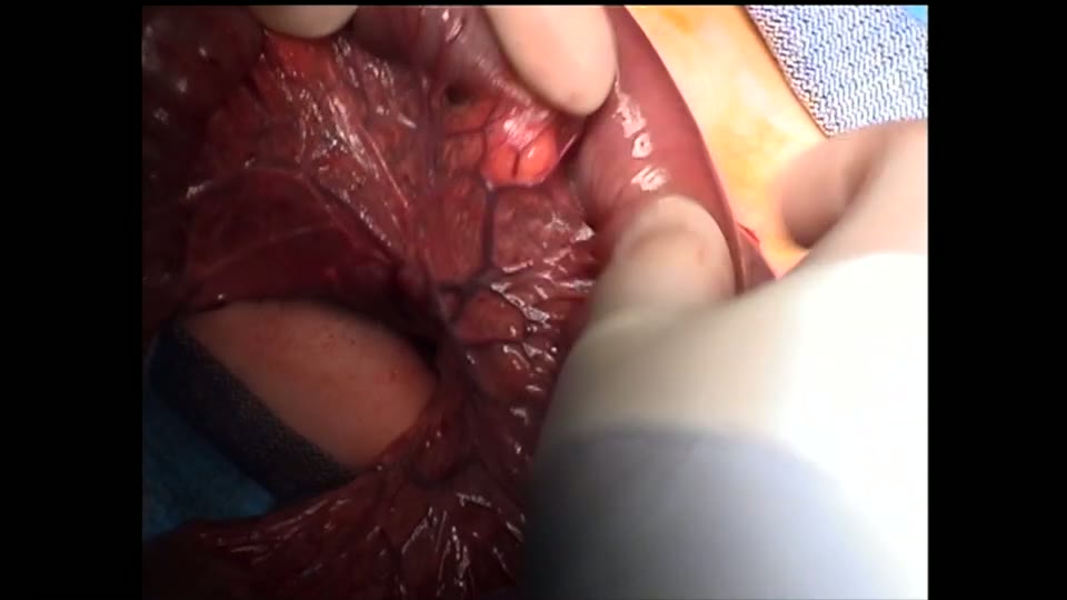
Open Bowel Anastomosis
Dr. Steve Rothenberg · Published May 2026
Video
Intestinal Atresia Types Explained: Grossfeld Classification for Pediatric Surgery
1 min · Published Sep 2026
Video
Derivation and validation of the Pediatric Community-Acquired Pneumonia Severity (PedCAPS) score: A prospective cohort study
50 s · Published Sep 2026
Video
FETO for Late-Diagnosed Severe Congenital Diaphragmatic Hernia (CDH) at Cincinnati Children's with Dr. Beth Rymeski
4 min · Published Sep 2026
Video
Severe Congenital Diaphragmatic Hernia (CDH) Case: FETO Management & Unexpected Findings with Dr. Beth Rymeski
3 min · Published Sep 2026
Video
Fetoscopic Endoluminal Tracheal Occlusion (FETO) Procedure: Step-by-Step Guide with Dr. Beth Rymeski
4 min · Published Sep 2026
Video
Choledochal Cyst Types Explained: Pediatric Surgery Fundamentals
1 min · Published Sep 2026
What the experts said
A 4-month-old, 5 kg infant with prenatally diagnosed CPAM underwent thoracoscopic left upper lobectomy.
An anterior approach was used with three ports: a 4 mm port in the posterior axillary line for the telescope and two 3 mm ports in the anterior axillary line, with the lower port later changed to 5 mm for the endoscopic clip applier.
A 3 mm sealer-dissector was used for the case.
The sealer was used to compress cysts in the left upper lobe to allow easier access to the pulmonary vessels.
The main trunk of the artery to the upper lobe was dissected out and sealed proximally and distally, then divided between the seals.
This technique (proximal and distal sealing before division) allows for a safe, effective, and reproducible method for sealing pulmonary vessels without risk of bleeding.
Each of the main branches of the superior pulmonary vein were individually isolated, dissected out, and sealed proximally and distally with division between the seals.
The major fissure was incomplete anteriorly.
The sealer was used to define the plane of the incomplete major fissure, and the lung between upper and lower lobes was sealed and divided.
A finger-fracture technique was used to divide the lung parenchyma and expose the artery as dissection continued posteriorly through the major fissure.
The superior and inferior branches of the pulmonary artery to the lingula were individually isolated, sealed, and divided using the 3 mm sealer.
The bronchus to the lingula was sealed with a 5 mm clip applier both proximally and distally and divided between the clips.
5 mm clips have proven to be an effective way to seal the bronchus in infants under 10 kg.
Each branch of the main bronchus to the upper lobe (apical-posterior and anterior) was individually sealed with a 5 mm clip and then divided.
The upper lobe was brought out through the lower trocar site in a piecemeal fashion.
The patient had a chest tube in for 24 hours and was discharged on the 2nd postoperative day.