19 views 0 likes

Dr. Steve Rothenberg

Pediatric Surgery · View profile →

Technique: Blinded Left Upper Lobectomy

Video Published 2019-01-11 Updated 2026-06-02

Timestops (10)

0:01
A 4 month old
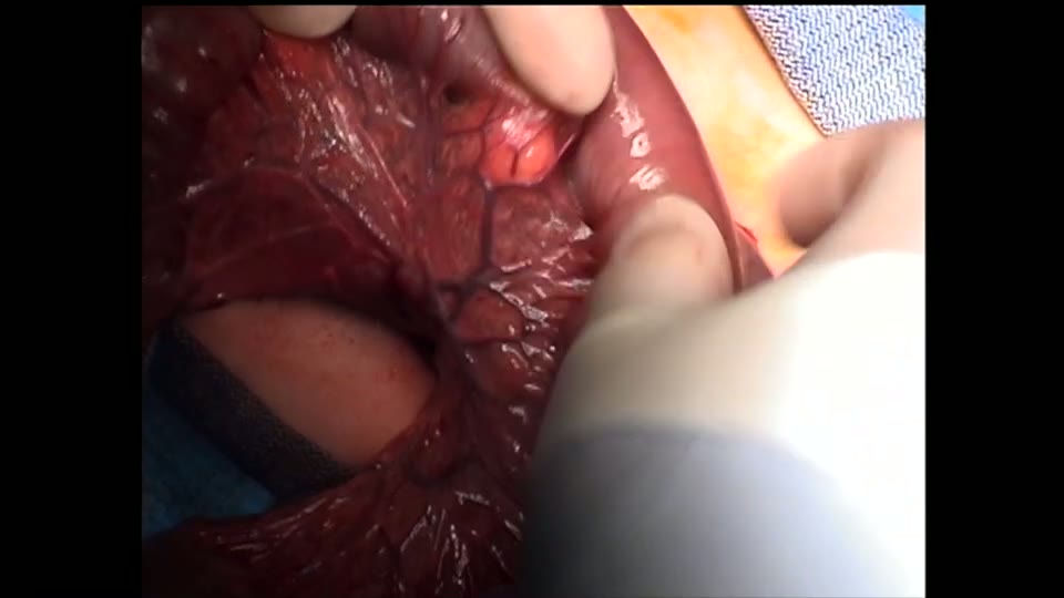
A 4 month old, 5 kg infant with a prenatally diagnosed CAm was brought to the OR for thoracoscopic left upper lobectomy.…
0:28
A new 3 millimeter sealer dissector was used for the case.
A new 3 millimeter sealer dissector was used for the case. Here you see the sealer being used to compress cysts in the l…
0:58
Here the main trunk of the artery to the upper lobe is disse…
Here the main trunk of the artery to the upper lobe is dissected out and sealed proximally and distally. And then divide…
1:25
With the main branches of the pulmonary artery to the apical…
With the main branches of the pulmonary artery to the apical posterior segment and anterior segment dissected out, atten…
2:04
With the main branches of the pulmonary vein to the upper lo…
With the main branches of the pulmonary vein to the upper lobe sealed, attention is turned to the major fissure. The maj…
2:24
As the section continues posteriorly towards the main pulmon…
As the section continues posteriorly towards the main pulmonary arteries, it courses through the major fissure, and almo…
2:54
The superior and inferior branches are individually isolated
The superior and inferior branches are individually isolated, sealed, and divided again using the 3 millimeter sealer. O…
3:25
5 millimeter clips have proven to be an effective way to sea…
5 millimeter clips have proven to be an effective way to seal. The bronchus in infants under 10 kg. With this done, the …
3:54
Each branch is individually sealed with a 5 millimeter clip.
Each branch is individually sealed with a 5 millimeter clip. And then divided proximal to this. With this done, the uppe…
4:25
Here you see the incisions at one month postoperatively.

Topic Overview

A single-speaker narration of a thoracoscopic left upper lobectomy in a 4-month-old, 5 kg infant with prenatally diagnosed congenital airway malformation (CAM). The procedure uses an anterior three-port approach with a 3 mm sealer-dissector for vessel and parenchymal division, and 5 mm clips for bronchial closure. The narrator describes sequential isolation and sealing of pulmonary artery branches, superior pulmonary vein branches, completion of the major fissure, and individual bronchial branches, followed by piecemeal specimen extraction. The patient was discharged on postoperative day 2.

Key Takeaways

  • 3mm sealer-dissector enables vessel division in infants without clips, reducing bleeding risk in small anatomy. (0:28)
  • 5mm clips effectively seal bronchi in infants <10kg; proven safe for bronchial closure in this weight class. (3:12)
  • Anterior 3-port approach with selective upsizing to 5mm allows instrument flexibility in 5kg infant thoracoscopy. (0:08)
  • Incomplete fissure managed by sealer-defining plane and finger-fracture technique to expose posterior vessels. (2:11)
  • Early discharge (POD 2) achievable after infant lobectomy with 24-hour chest tube protocol. (4:13)

Inside this episode

Kai, the Library's AI content creator, listened to this episode and mapped who's speaking, the chapters, key claims, and cases. Every item links to the exact moment in the recording.

AI-enriched

Who's speaking

  • Speaker 1

Chapters

  • 0:01Patient and Port Placement — Introduction of a 4-month-old, 5 kg infant with prenatal CAM diagnosis. Anterior approach with three ports: 4 mm posterior axillary for telescope, two 3 mm anterior axillary (lower later changed to 5 mm for clip applier). A 3 mm sealer-dissector is used to decompress cystic lung tissue.
  • 0:43Pulmonary Artery and Vein Division — Upper lobe retracted inferiorly to expose superior pulmonary artery branches (apical posterior, anterior). Main trunk and branches sealed proximally and distally, then divided. Superior pulmonary vein branches individually isolated, sealed, and divided in the same manner.
  • 2:04Major Fissure and Lingular Vessels — Incomplete major fissure defined and divided with sealer. Finger-fracture technique used posteriorly to expose artery. Small posterior arterial branch identified. Lingular artery branches (superior and inferior) individually isolated, sealed, and divided with 3 mm sealer.
  • 3:16Bronchial Division and Specimen Extraction — Lingular bronchus sealed with 5 mm clips proximally and distally, then divided. Main upper lobe bronchus visualized at its bifurcation; apical posterior and anterior branches individually clipped with 5 mm clips and divided. Upper lobe extracted piecemeal through lower trocar site. Chest tube removed at 24 hours, discharge on postoperative day 2.

Key claims

  • 0:01A 4-month-old, 5 kg infant with prenatally diagnosed CAM underwent thoracoscopic left upper lobectomy. — Speaker 1
  • 0:08An anterior approach was used with three ports: 4 mm in the posterior axillary line for the telescope, and two 3 mm ports in the anterior axillary line. — Speaker 1
  • 0:16The lower 3 mm port was later changed to 5 mm for access of the endoscopic clip applier. — Speaker 1
  • 0:28A new 3 mm sealer-dissector was used for the case. — Speaker 1
  • 0:32The sealer was used to compress cysts in the left upper lobe to allow easier access to the pulmonary vessels. — Speaker 1
  • 0:46The upper lobe was retracted inferiorly to expose the superior branches of the pulmonary artery (apical posterior and anterior branches). — Speaker 1
  • 0:58The main trunk of the artery to the upper lobe was dissected out and sealed proximally and distally, then divided between the seals. — Speaker 1
  • 1:10This technique allows for a safe, effective, and reproducible method for sealing pulmonary vessels without risk of bleeding. — Speaker 1
  • 1:36Each of the main branches of the superior pulmonary vein were individually isolated, dissected out, and then sealed proximally and distally with division of the vessel between the seals. — Speaker 1
  • 2:11The major fissure was incomplete anteriorly. — Speaker 1
  • 2:11The sealer was used to help define the plane of the incomplete major fissure, and the lung between the upper and lower lobes was sealed and then divided. — Speaker 1
  • 2:24As dissection continued posteriorly towards the main pulmonary arteries through the major fissure, an almost finger-fracture technique was used to divide the lung parenchyma and expose the artery. — Speaker 1
  • 2:40A small posterior branch of the artery going to the upper lobe was identified. — Speaker 1
  • 2:54The superior and inferior branches of the pulmonary artery going to the lingula were individually isolated, sealed, and divided using the 3 mm sealer. — Speaker 1
  • 3:12The bronchus to the lingula was sealed with a 5 mm clip applier, both proximally and distally, and divided between these. — Speaker 1
  • 3:255 mm clips have proven to be an effective way to seal the bronchus in infants under 10 kg. — Speaker 1
  • 3:38The main bronchus to the remainder of the upper lobe was visualized at its bifurcation between the apical posterior and anterior branches. — Speaker 1
  • 3:54Each bronchial branch was individually sealed with a 5 mm clip and then divided proximal to this. — Speaker 1
  • 4:05The upper lobe was brought out through the lower trocar site in a piecemeal fashion. — Speaker 1
  • 4:13The child had a chest tube in for 24 hours and was discharged on the 2nd postoperative day. — Speaker 1

Cases discussed

  • 0:014-month-old, 5 kg infant with prenatally diagnosed congenital airway malformation (CAM) of the left upper lobe.
This episode was analyzed and enriched by Kai, the Library's AI content creator. Every item links to the moment it comes from — click a timestamp to listen in context.
Written for:

Thoracoscopic Lobectomy in Infants: Vessel Sealing Without Staplers

The episode's main topic retold as a plain-language walkthrough — what it is, why it matters, and what the speakers concluded. Written by Kai from the episode transcript and reviewed before publishing.

For the care team · Explainer · AI-written, human-reviewed

Why This Approach Exists

Pediatric thoracoscopic lobectomy confronts a problem that does not exist in adult surgery: the chest wall is compliant, the working space is measured in centimeters, and endoscopic staplers — the workhorse of adult VATS — are often too large for the hilar structures of an infant. A 4-month-old with a 5 kg body weight cannot accommodate a stapler cartridge across a 3 mm pulmonary artery branch 0:01. The alternative is individual vessel ligation, which is tedious and carries its own bleeding risk when sutures slip in small, fragile tissue. The technique demonstrated here — sequential isolation and energy-based sealing of each named vessel and bronchus — offers a middle path: no staplers, no ties, and reproducible hemostasis in a confined space.

The Core Problem

Congenital pulmonary airway malformation (CAM, formerly CCAM) is the most common indication for neonatal lobectomy. The lesion is typically cystic, bulky, and distorts normal hilar anatomy. In this case, a prenatally diagnosed CAM occupied the left upper lobe of a 4-month-old infant 0:01. The surgical goal is complete lobectomy with minimal blood loss, no air leak, and preservation of the remaining lung. The challenge is that the cystic mass obscures the pulmonary artery branches, the fissure is often incomplete, and the margin for error in a 5 kg child is narrow.

How the Approach Works

Port Placement and Access

The anterior approach uses three ports: a 4 mm posterior axillary port for the telescope and two 3 mm anterior axillary ports, one of which is later upsized to 5 mm to accommodate a clip applier [c2, c3]. This configuration keeps instruments anterior to the hilum and avoids crowding. A 3 mm sealer-dissector — a bipolar energy device that both coagulates and divides tissue — is the primary working instrument 0:28.

Cyst Decompression

Before hilar dissection begins, the sealer is used to compress the cystic lung tissue 0:32. This is not cautery for hemostasis; it is mechanical decompression to collapse the mass and improve visualization of the vessels beneath. In a small chest, reducing the bulk of the specimen changes the operative field.

Arterial Division

The upper lobe is retracted inferiorly to expose the superior branches of the left pulmonary artery — the apical posterior and anterior segmental branches 0:46. Each vessel is individually dissected free, sealed proximally and distally with the energy device, then divided between the seals 0:58. The discussant describes this as a safe, effective, and reproducible method for sealing pulmonary vessels without risk of bleeding 1:10. The key is double sealing: proximal and distal application before division eliminates the open vessel end that would otherwise require a clip or tie.

Venous Division

The superior pulmonary vein branches are handled identically: individual isolation, proximal and distal sealing, division between seals 1:36. The sequence — artery first, then vein — is standard, but the technique is the same for both.

Fissure Management

The major fissure in this case was incomplete anteriorly 2:11, meaning the upper and lower lobes were not fully separated by a natural cleavage plane. The sealer was used to define the fissure plane and divide the parenchymal bridge 2:11. Posteriorly, where the fissure approached the main pulmonary artery, the discussant used an "almost finger-fracture technique" — blunt dissection to spread the lung tissue and expose the artery without cutting blindly 2:24. A small posterior arterial branch to the upper lobe was identified and divided 2:40.

Lingular Vessels and Bronchus

The lingula — the tongue-like projection of the left upper lobe — has its own arterial supply. The superior and inferior lingular artery branches were individually isolated, sealed, and divided with the 3 mm sealer 2:54. The lingular bronchus, however, was not sealed with energy. Instead, it was clipped proximally and distally with a 5 mm clip applier and divided between the clips 3:12. The discussant notes that 5 mm clips have proven to be an effective way to seal the bronchus in infants under 10 kg 3:25. This is a weight-based threshold: clips are reliable in small airways where staplers do not fit, but energy sealing of the bronchus risks thermal injury to the adjacent airway.

Main Bronchial Division

The main upper lobe bronchus was visualized at its bifurcation into apical posterior and anterior branches 3:38. Each branch was clipped with a 5 mm clip and divided proximal to the clip 3:54. The upper lobe was then extracted piecemeal through the lower trocar site 4:05.

Outcome

The child was extubated in the operating room, had a chest tube for 24 hours, and was discharged on postoperative day 2 4:13. This is a benchmark outcome for uncomplicated infant lobectomy.

When to Involve Pediatric Thoracic Surgery

CAM diagnosed prenatally or in early infancy warrants referral if the lesion is symptomatic (recurrent infection, respiratory distress) or if imaging shows a dominant cyst or solid component that raises concern for malignancy. Asymptomatic lesions are increasingly managed with observation, but the threshold for resection is lower in infants than in older children because the risk of infection is higher and the lung has greater capacity for compensatory growth. Timing is typically in the first months of age, after the neonatal period but before the child outgrows the window for minimally invasive resection. If you are managing a prenatal CAM diagnosis, involve pediatric surgery early in the third trimester to plan postnatal imaging and timing of intervention.

Takeaways from this story

  • In infants under 10 kg, 5 mm clips reliably seal bronchi where staplers are too large for hilar structures.
  • Energy-based vessel sealing with proximal and distal application before division eliminates bleeding risk without ties or staplers.
  • Cyst decompression with the sealer before hilar dissection improves visualization in bulky CAM lesions.
  • Incomplete fissures require combined energy sealing anteriorly and blunt finger-fracture dissection posteriorly to safely expose the artery.

Keywords

Hashtags

Transcript

Comments

Loading comments…