19 views 0 likes

StayCurrentMD

GCMD Space · View profile →

Spontaneous Pneumothorax: Lung Lesions

Video Published 2019-01-11 Updated 2022-08-22

Timestops (5)

Topic Overview

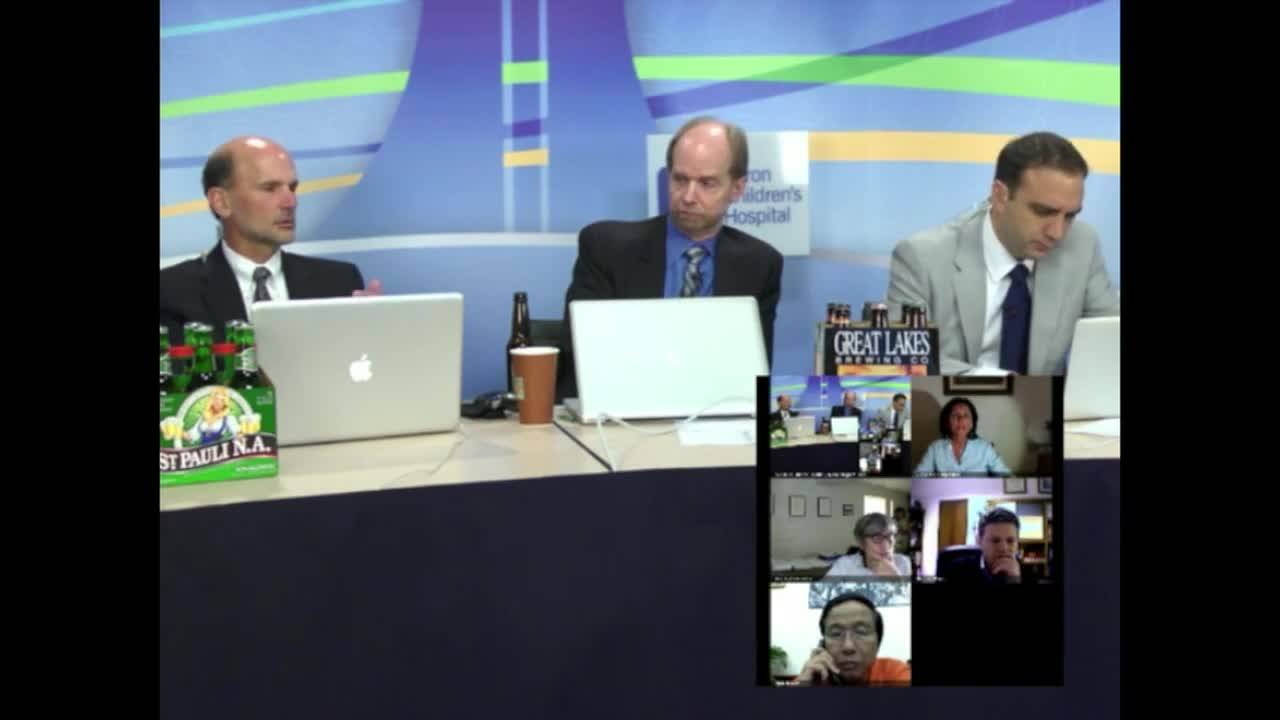
A multidisciplinary panel discussion on the management of primary spontaneous pneumothorax in pediatric patients, focusing on initial treatment decisions, the role of CT imaging, and surgical approaches for bleb disease. The discussants debate observation versus chest tube placement for a 14-year-old with 20% pneumothorax, the timing and utility of CT scans, and whether to perform video-assisted thoracoscopic surgery (VATS) at first presentation or wait for recurrence. Key points of disagreement include whether to treat blebs prophylactically when found on imaging or thoracoscopy, whether to address bilateral bleb disease in one or two stages, and optimal pleurodesis techniques (mechanical apical pleurectomy versus chemical talc pleurodesis).

Key Takeaways

  • 50% recurrence after chest tube alone; 75% after second event—consider early VATS for high-risk patients (7:27)
  • CT poorly predicts bleb disease vs thoracoscopy; nearly all spontaneous pneumothorax patients have blebs on direct visualization (13:00)
  • Chemical talc pleurodesis shows shorter OR time and lower recurrence than mechanical pleurectomy but may complicate future chest access (19:08)
  • Apical pleurectomy limited to third intercostal space causes more postop pain but preserves future surgical access (19:49)

Inside this episode

Kai, the Library's AI content creator, listened to this episode and mapped who's speaking, the chapters, key claims, and cases. Every item links to the exact moment in the recording.

AI-enriched

Who's speaking

  • Mark — guest
  • Todd — host
  • Marcello — guest
  • Giovanna — guest
  • Liam — guest

Chapters

  • 0:00Case Presentation and Initial Management Approaches — Mark presents a 14-year-old with acute left-sided chest pain, shortness of breath, and 20% pneumothorax. Panel discusses initial management options ranging from observation to immediate VATS. Regional practice variation emerges: Argentina favors chest tube in ED, Italy/Japan favor observation, while Mark's center takes patients to OR for chest tube under anesthesia, which then facilitates diagnostic thoracoscopy.
  • 4:36Anesthesia Approach and Rationale for Early VATS — Discussion of whether chest tubes are placed under local anesthesia at bedside versus general anesthesia in OR. Mark argues that if a child requires general anesthesia for chest tube placement, it makes sense to perform diagnostic thoracoscopy simultaneously to identify blebs, given 50% recurrence rates. Todd challenges whether presence of blebs necessarily predicts recurrence, noting lack of studies separating bleb-positive from bleb-negative patients in terms of recurrence risk.
  • 8:41Role and Timing of CT Imaging — Debate over CT scan utility in spontaneous pneumothorax. Liam advocates CT scan on stable patients before intervention. Mark notes CT can reveal extent of disease including contralateral blebs and lower lobe involvement, helping inform patient/family decisions especially for high-risk activities. Todd questions whether CT changes management since he treats based on clinical recurrence. Alan cites recent study from Kansas City showing poor CT sensitivity for bleb detection compared to thoracoscopy. Panel agrees most patients likely have bleb disease whether detected or not.
  • 15:17Management of Bilateral Bleb Disease — Discussion of approach when CT or thoracoscopy reveals bilateral blebs. Mark and Giovanna favor treating both sides simultaneously to prevent contralateral recurrence. Todd, Marcello, and Yama treat only the symptomatic side. Giovanna reports experiencing immediate postoperative contralateral recurrence after treating only symptomatic side, which changed her practice. Audience poll shows 70% would treat symptomatic side only. This debate reinforces the argument for preoperative CT imaging if bilateral treatment is being considered.
  • 19:03Pleurodesis Techniques and Recurrence Prevention — Panel discusses mechanical versus chemical pleurodesis methods. Todd describes evolution from bovie pad abrasion to apical pleurectomy (taught by Steve) to aerosolized talc, noting pleurectomy caused more postoperative pain. Steve advocates apical pleurectomy limited to 2-3 rib spaces, avoiding talc due to concern about future chest access. Mark describes hydrostatic pleurectomy technique using suction irrigator to dissect entire hemithorax pleura. Steve reveals he ligates the lung apex when no blebs are visible, then performs apical pleurectomy. Consensus that recurrence rates remain significant even after VATS with blebectomy and pleurodesis.

Key claims

  • 7:2750% of patients who undergo simple chest tube placement for symptomatic pneumothorax will have recurrence — Mark
  • 7:55Second recurrence rate is approximately 75% — Mark
  • 5:12Most spontaneous pneumothorax patients are adolescents around 18 years old — Marcello
  • 13:00A recent study from Saint Peter in Kansas City found CT scan was very poor at defining bleb disease compared to subsequent thoracoscopy — Mark
  • 13:17Almost all spontaneous pneumothorax patients have some degree of bleb disease whether defined early or not — Mark
  • 13:28Bleb disease doesn't necessarily mean recurrence — Mark
  • 14:44Patients with spontaneous pneumothorax usually don't present in extremis, they usually come in with pain — Mark
  • 19:08More people are using chemical pleurodesis instead of mechanical pleurodesis and finding better results, shorter OR times, and decreased recurrence rates — Mark
  • 19:17There is still a pretty high recurrence rate even after VATS with blebectomy and mechanical and/or chemical pleurodesis — Mark
  • 19:49Apical pleurectomy causes more postoperative pain than talc pleurodesis — Todd
  • 20:21Talc creates random massive chemical pleurodesis throughout the chest, which may complicate future chest access — Todd
  • 21:48Apical pleurectomy is limited to the apex of the lung, typically down to the third intercostal space when apical blebs are seen — Todd
  • 11:43CT scan can reveal blebs in the superior segment of the lower lobe and contralateral side, not just upper lobe — Mark
  • 9:04In Argentina, chest tubes for pneumothorax are typically placed as thin double pigtail catheters attached to Heimlich valve under local anesthesia — Marcello
  • 20:48Hydrostatic pleurectomy technique involves making a small pleural incision, inserting suction irrigator, sealing pleura around it, and irrigating to dissect entire pleura which can then be rolled up — Mark

Cases discussed

  • 0:4214-year-old male with acute onset left-sided chest pain and shortness of breath, found to have approximately 20% left pneumothorax
  • 12:11Female patient with spontaneous pneumothorax going on 6-week wilderness trip, found to have bilateral blebs on CT scan
  • 18:24Patient with bilateral bleb disease currently hospitalized in Argentina

Points of disagreement

  • 6:23Whether to perform VATS at first presentation versus waiting for recurrence
    • Mark: If child requires general anesthesia for chest tube, should perform diagnostic thoracoscopy and treat blebs at first presentation given 50% recurrence rate
    • Todd: Lack of evidence that presence of blebs predicts recurrence; can treat at second recurrence when 75% will recur; questions doing potentially unnecessary procedures on 50% of patients
  • 8:41Role and timing of CT scan in spontaneous pneumothorax
    • Liam: Get CT scan on all stable patients before intervention to know the nature and cause of pneumothorax
    • Mark: CT scan useful to define extent of disease including contralateral blebs and help inform patient/family decisions
    • Todd: CT scan doesn't change management; treats based on clinical recurrence regardless of imaging findings
  • 17:05Management of bilateral bleb disease - one stage versus two stage
    • Mark: Would do both sides at same time if bleb disease seen bilaterally
    • Giovanna: Do bilateral because had experience with immediate postoperative contralateral recurrence after treating only symptomatic side
    • Liam: Would do bilateral
    • Todd: Do symptomatic side only
    • Marcello: Treat symptomatic side only; if contralateral side is asymptomatic on CT, send home with observation
  • 19:31Pleurodesis technique - mechanical versus chemical
    • Todd: Previously used apical pleurectomy but now uses aerosolized talc for ease and less postoperative pain
    • Todd: Performs apical pleurectomy limited to 2-3 rib spaces; avoids talc due to concern about creating adhesions throughout chest that would complicate future chest access
    • Mark: Uses hydrostatic pleurectomy technique; agrees talc is probably better but shares concern about future chest access; saves talc for recurrences after other approaches

Open questions

  • What is the recurrence rate specifically for patients with blebs versus those without blebs after first spontaneous pneumothorax?
  • How sensitive is CT scan for detecting blebs compared to thoracoscopy?
  • What is the optimal pleurodesis technique balancing recurrence prevention with postoperative pain and future chest access?
  • Should bilateral bleb disease be treated in one stage or two stages?
  • What is the true recurrence rate after VATS with blebectomy and pleurodesis?
  • How far should apical pleurectomy extend to be effective while minimizing complications?
This episode was analyzed and enriched by Kai, the Library's AI content creator. Every item links to the moment it comes from — click a timestamp to listen in context.
Written for:

Primary Spontaneous Pneumothorax: When to Intervene and How Aggressively

The episode's main topic retold as a plain-language walkthrough — what it is, why it matters, and what the speakers concluded. Written by Kai from the episode transcript and reviewed before publishing.

For the care team · Explainer · AI-written, human-reviewed

Why This Exists as a Distinct Problem

Primary spontaneous pneumothorax — lung collapse without trauma or underlying disease — occurs overwhelmingly in otherwise healthy adolescents, typically around 18 years old 5:12. The clinical presentation is usually acute chest pain and shortness of breath, not respiratory distress 14:44. The immediate management question is straightforward: observation, chest tube, or surgery. The strategic question is harder: how aggressively should you treat the first event to prevent recurrence, and does identifying blebs on imaging or thoracoscopy change that calculus?

The Core Clinical Problem

The recurrence rate after conservative management — observation or chest tube alone — approaches 50% 7:27. After a second event, recurrence climbs to approximately 75% 7:55. These are not trivial numbers for an adolescent whose life revolves around school, sports, and independence. The debate centers on whether the presence of blebs — small subpleural air pockets visible on CT or thoracoscopy — predicts recurrence strongly enough to justify definitive intervention at first presentation, or whether recurrence itself is the appropriate trigger for surgery.

The discussants frame this tension clearly: if you scope every first pneumothorax and resect every bleb you find, you may be operating on half the patients unnecessarily. If you wait for clinical recurrence, you subject half the patients to a second event that could have been prevented.

How the Approach Works

Initial management varies by region and institutional culture. In Argentina, thin double-pigtail catheters attached to Heimlich valves are placed under local anesthesia in the emergency department 9:04. In other centers, adolescents and their families prefer general anesthesia, which shifts the procedure to the operating room. One discussant argues that if the child is already under anesthesia for chest tube placement, diagnostic thoracoscopy adds minimal burden and allows immediate treatment of visible blebs [q2]. The counterargument: you are basing intervention on anatomic findings — blebs — whose predictive value for recurrence is unproven when separated from clinical recurrence itself [q4].

CT imaging is contested. It can reveal blebs in the contralateral lung and the superior segment of the lower lobe, not just the upper lobe apex 11:43. For a patient planning high-risk activities — wilderness trips, remote travel — this information may inform shared decision-making even if it does not change the surgical plan. But a recent study from Kansas City found CT poorly sensitive for bleb detection compared to thoracoscopy 13:00, and most discussants believe nearly all spontaneous pneumothorax patients have some degree of bleb disease whether imaged or not 13:17. The critical point: bleb disease does not necessarily predict recurrence 13:28.

When surgery is performed, the technical approach includes resection of visible blebs and some form of pleurodesis to promote adhesion between lung and chest wall. Chemical pleurodesis with aerosolized talc is increasingly favored for shorter operative times and potentially lower recurrence rates 19:08, though recurrence remains significant even after video-assisted thoracoscopic surgery (VATS) with blebectomy and pleurodesis 19:17. Mechanical pleurodesis by apical pleurectomy — stripping the pleura from the chest wall apex down to approximately the third intercostal space 21:48 — causes more postoperative pain than talc 19:49 but avoids the concern that diffuse talc pleurodesis may complicate future chest access 20:21. One discussant describes hydrostatic pleurectomy: making a small pleural incision, inserting a suction irrigator, sealing around it, and using irrigation to dissect the pleura, which can then be removed 20:48.

Where Practice is Genuinely Contested

The bilateral bleb question divides the panel cleanly. If CT or thoracoscopy reveals blebs on both sides, should you treat the asymptomatic contralateral lung? One discussant changed practice after experiencing immediate postoperative contralateral recurrence when treating only the symptomatic side [q13]. Others treat only the symptomatic side, reasoning that prophylactic contralateral intervention exposes the patient to unnecessary risk. An audience poll showed 70% would treat the symptomatic side only.

The deeper disagreement is whether visible blebs at first presentation justify intervention or whether clinical recurrence is the appropriate threshold. No study has separated first-event patients into bleb-positive and bleb-negative cohorts and compared recurrence rates between them [q5]. Without that data, the choice between early definitive surgery and watchful waiting rests on how you weight the burden of a second event against the risk of unnecessary surgery.

When to Involve This Team

Refer at first presentation if the pneumothorax is large, symptomatic, or the patient is engaged in high-risk activities where recurrence would be dangerous. Refer urgently for any pneumothorax with respiratory compromise, though true extremis is rare 14:44. Refer after first recurrence — at that point, the 75% risk of third recurrence 7:55 makes definitive intervention the standard approach. If a primary-care provider or emergency physician places a chest tube and the lung re-expands, pediatric surgery should be involved in discharge planning and recurrence counseling, because half these patients will return 7:27.

Takeaways from this story

  • 50% of adolescents with first spontaneous pneumothorax will have recurrence after conservative management alone
  • CT scan sensitivity for blebs is poor compared to thoracoscopy, and most patients likely have some bleb disease regardless
  • Presence of blebs does not reliably predict recurrence — no study has separated bleb-positive from bleb-negative first events
  • Apical pleurectomy limited to 2-3 rib spaces causes more pain than talc but avoids diffuse pleural adhesions

Keywords

Hashtags

Transcript

Comments

Loading comments…