18 views 0 likes

StayCurrentMD

GCMD Space · View profile →

Physiology: Pectus Innovations

Video Published 2019-01-11 Updated 2022-08-22

Timestops (6)

Topic Overview

A multidisciplinary discussion of pectus excavatum physiology and management, focusing on cardiopulmonary exercise testing and echocardiographic findings in older patients. The speakers present data showing 24-30% improvement in cardiac output after repair, with TEE revealing right ventricular compression and improved flow velocities post-operatively. Key debates include optimal surgical timing (young children vs. adolescents), the role of vacuum bell therapy, genetic evaluation in connective tissue disorders, and counseling asymptomatic adolescents with severe deformity given the risk of future symptom development.

Key Takeaways

  • TEE reveals RV compression in pectus: post-op cardiac output improves 24-30%, with triple RV outflow velocity vs pre-op. (2:12)
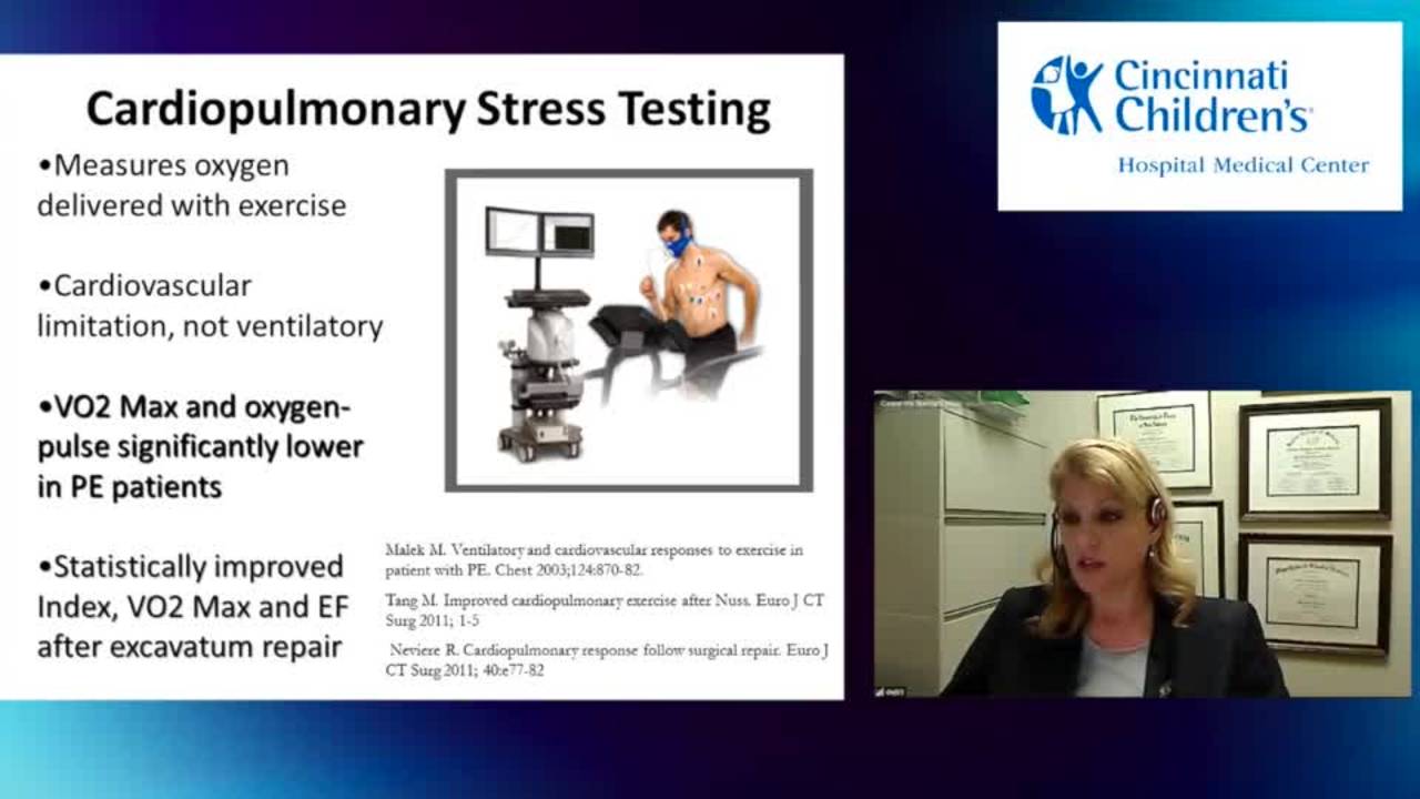
  • Cardiopulmonary exercise testing with VO2 max is ideal for pectus evaluation, showing flat stroke volume curves vs normal. (5:13)
  • Transthoracic echo misses RV pathology in 99% of pectus patients due to inadequate windows for right heart visualization. (1:24)
  • Minimally invasive repair shows superior cardiac output gains (30%) vs open repair, likely due to mixed pathology in open cases. (3:57)
  • No RCTs exist comparing repaired vs non-repaired pectus patients; selection bias limits evidence as most clinic patients seek surgery. (19:31)

Inside this episode

Kai, the Library's AI content creator, listened to this episode and mapped who's speaking, the chapters, key claims, and cases. Every item links to the exact moment in the recording.

AI-enriched

Who's speaking

  • Speaker 1 — guest
  • Speaker 2 — host
  • Speaker 3 — guest
  • Speaker 4 — host
  • Speaker 5 — guest
  • Speaker 6 — guest

Chapters

  • 0:00Cardiopulmonary Exercise Testing and Echocardiographic Findings — Presentation of diagnostic workup for older pectus patients, including TEE findings showing right ventricular compression, velocity improvements, and cardiac output increases of 24-30% post-repair. Discussion of VO2 max testing showing flat stroke volume curves in pectus patients that normalize after repair.
  • 7:00Cardiologist Perspective on Exercise Testing — Cardiology discussion of exercise testing utility in pectus patients, noting it is used extensively in congenital heart disease but less commonly for pectus alone, with potential value in borderline cases.
  • 8:32Genetic Considerations: Ehlers-Danlos Type 4 — Discussion of surgical approach to Ehlers-Danlos type 4 (vascular type), emphasizing tissue fragility concerns, need for genetic confirmation, and patient confusion between hypermobile and vascular subtypes based on internet research.
  • 10:52Optimal Surgical Timing Debate — Debate over surgical timing, weighing chest wall flexibility and pain tolerance in younger children against growth disturbance risk and bar outgrowth. Discussion of geographic variation (Korea operates at ages 3-5, US trend toward older patients) and lack of definitive data.
  • 15:41Vacuum Bell Therapy and Young Children — Discussion of vacuum bell as non-operative option for young children, noting variable patient compliance and effectiveness dependent on chest flexibility and consistent use.
  • 18:51Counseling Asymptomatic Adolescents — Approach to counseling asymptomatic adolescents with severe deformity, acknowledging selection bias in symptomatic adult populations, presenting physiologic data, and allowing patient choice while noting high likelihood of future symptom development.

Key claims

  • 1:2499% of pectus patients receive transthoracic echos reported as normal with no issues — Speaker 1
  • 1:32Transthoracic echo has difficulty obtaining adequate windows to visualize the right side of the heart — Speaker 1
  • 2:12TEE shows nearly triple the velocity or volume going through the right ventricular outflow tract post-operatively — Speaker 1
  • 2:25Left ventricular flow also shows significant improvement when right ventricular flow increases, because the heart functions as a pump — Speaker 1
  • 2:59Cardiac output can improve from 55% (normal range) to 77% in older pectus patients after repair — Speaker 1
  • 3:26Heart contractility, synchrony, and mobility show significant differences between pre-op and post-op in pectus patients — Speaker 1
  • 3:57In 168 patients studied pre and post-op, the MIRP (minimally invasive) group showed 30% increase in cardiac output — Speaker 1
  • 4:24All pectus patients together showed 24% increase in cardiac output post-operatively — Speaker 1
  • 3:57Open repair group does not improve cardiac output as much as minimally invasive group, likely because open cases include mixed pathology like malunion — Speaker 1
  • 4:53Standard Bruce protocol ischemic stress testing is not the appropriate test for pectus patients unless ischemia is suspected — Speaker 1
  • 5:13Cardiopulmonary exercise testing with VO2 measurement separates cardiac efficiency from pulmonary component, making it ideal for pectus evaluation — Speaker 1
  • 5:27VO2 max and anaerobic VO2 are very abnormal in pectus patients while other parameters remain normal — Speaker 1
  • 5:39A French study of 125 adults showed cardiopulmonary exercise parameters statistically improve and normalize after surgical pectus repair — Speaker 1
  • 6:02Pectus patients show limited ability to increase stroke volume during exercise, initially compensating with heart rate but then plateauing when volume increase is needed due to chest cage restriction — Speaker 1
  • 6:19Pectus patients show flat stroke volume curves during exercise testing while normal patients show upward slopes that peak at maximum — Speaker 1
  • 7:23Exercise testing is used 5-6 times daily in congenital heart disease clinic for decision-making about palliation and conduit replacement — Speaker 3
  • 7:39Exercise testing has not been used extensively for pectus patients alone in the cardiology practice described — Speaker 3
  • 9:03Ehlers-Danlos type 4 (vascular type) does not typically present with pectus excavatum as it is not primarily a skeletal or hypermobile disorder — Speaker 5
  • 9:21Ehlers-Danlos type 4 involves internal tissue fragility requiring specially managed repairs — Speaker 5
  • 10:09Visible veins and family history of abdominal aortic aneurysm do not indicate Ehlers-Danlos vascular type — Speaker 5
  • 14:51Younger children have more pliable chest walls, allowing repair with less force (described as 'popsicle stick' level) — Speaker 1
  • 15:01Adults have tremendous pain from pectus repair because their chest walls are rigid — Speaker 1
  • 15:06In Korea, pectus repairs are performed on 3-5 year olds with very small bar sets — Speaker 1
  • 15:18The trend in the United States has moved from repairing young patients to operating on older populations — Speaker 1
  • 15:41Donald Nuss initially performed pectus repairs on very young patients, as young as 3 years of age — Speaker 2
  • 16:37The Nuss institution with thousands of patients has moved away from operating on young children — Speaker 1
  • 16:57Korean surgeons repair pectus in young children routinely and report no problems, considering it unusual to wait — Speaker 1
  • 17:52Vacuum bell works best with very flexible chest walls and requires religious compliance — Speaker 1
  • 18:03Patient compliance with vacuum bell varies greatly, with some children only tolerating 10 minutes of use despite bribing — Speaker 1
  • 19:31There are no randomized controlled trials separating pectus patients who received repair from those who did not — Speaker 1
  • 19:39Selection bias exists in surgical practice because most patients seen are symptomatic, while thousands of asymptomatic adults with pectus may exist — Speaker 1
  • 20:2595% of patients presenting to the pectus clinic are seeking surgery — Speaker 1

Points of disagreement

  • 10:52Optimal age for pectus repair
    • Speaker 1: Favors adolescent repair (12-13+) due to concerns about bar outgrowth and growth disturbance in young children, though acknowledges severely symptomatic young children may need earlier repair
    • Speaker 4: Previously opposed to repair before age 12-13, but being convinced by arguments for younger repair based on pain tolerance and chest wall flexibility
    • Speaker 2: Questions whether delaying surgery increases narcotic exposure and addiction risk in older patients who require more pain control
  • 7:04Value of exercise testing in pectus patients
    • Speaker 1: Considers cardiopulmonary exercise testing critical for insurance approval and documenting physiologic impairment in pectus patients
    • Speaker 3: Uses exercise testing extensively in congenital heart disease but has not found it necessary for pectus patients, as surgical decisions are made on other bases

Open questions

  • What is the true natural history of untreated pectus excavatum in asymptomatic patients?
  • Does early surgical repair in young children increase risk of bar outgrowth and need for revision?
  • Does delaying pectus repair to older ages increase narcotic exposure and addiction risk?
  • Why did the Nuss institution shift away from operating on young children after initially reporting good results?
  • What is the optimal age for pectus repair balancing chest wall flexibility, pain tolerance, growth concerns, and long-term outcomes?
  • How can vacuum bell compliance be improved to make it a more reliable non-operative option?
This episode was analyzed and enriched by Kai, the Library's AI content creator. Every item links to the moment it comes from — click a timestamp to listen in context.
Written for:

Pectus Excavatum as Cardiopulmonary Disease: Physiologic Evidence Beyond Cosmesis

The episode's main topic retold as a plain-language walkthrough — what it is, why it matters, and what the speakers concluded. Written by Kai from the episode transcript and reviewed before publishing.

For the care team · Explainer · AI-written, human-reviewed

Why This Exists as a Discipline

Pectus excavatum — the inward depression of the anterior chest wall — was long dismissed as a cosmetic problem. Patients complained of exercise intolerance and dyspnea; standard transthoracic echocardiography came back normal; surgeons operated primarily on adolescents seeking body-image correction. The emergence of minimally invasive repair techniques and advanced cardiac imaging has forced a reckoning: this is measurable cardiopulmonary pathology, not vanity surgery. The question is no longer whether pectus affects the heart, but how much impairment justifies intervention and at what age.

The Core Problem

The depressed sternum compresses the right ventricle and displaces the heart leftward. Standard transthoracic echo reports 99% of these patients as normal because acoustic windows cannot adequately visualize the compressed right heart 1:24 1:32. Transesophageal echo reveals the truth: right ventricular outflow tract velocities nearly triple after surgical correction, and left ventricular output improves in tandem because the heart functions as a pump — right-sided flow directly drives left-sided performance 2:12 2:25. In older patients, cardiac output can jump from 55% to 77% post-repair 2:59. The chest cage physically restricts cardiac filling and ejection.

The functional consequence appears most clearly on cardiopulmonary exercise testing with VO2 measurement. Normal individuals increase stroke volume progressively during exertion, producing an upward-sloping curve that peaks at maximum effort. Pectus patients compensate initially by raising heart rate, but when further cardiac output is needed, they cannot increase stroke volume due to chest cage restriction — the curve flattens 6:02 6:19. VO2 max and anaerobic threshold are abnormal while other parameters remain normal, isolating the cardiac efficiency deficit from pulmonary mechanics 5:27. A French study of 125 adults demonstrated statistical normalization of these parameters after surgical repair 5:39.

How the Approach Works

The diagnostic workup for older or symptomatic patients now includes:

  • Transesophageal echocardiography to visualize right ventricular compression, chamber dimensions, and flow velocities pre- and post-operatively. Three-dimensional imaging reveals improvements in contractility, synchrony, and myocardial mobility that standard views miss 3:26.
  • Cardiopulmonary exercise testing — not the standard Bruce protocol ischemic stress test, which is appropriate only if coronary disease is suspected 4:53. VO2 measurement separates cardiac efficiency from pulmonary mechanics, making it ideal for pectus evaluation 5:13. Insurance resistance to post-operative testing in the United States limits longitudinal data, but European centers have documented normalization after repair.

In a series of 168 patients studied pre- and post-operatively, minimally invasive repair produced a 30% increase in cardiac output; all pectus patients together showed a 24% increase 3:57 4:24. Open repair cases — which include mixed pathology like sternal malunion — showed smaller gains 3:57.

Congenital heart disease clinics use exercise testing routinely to guide decisions about palliation and conduit replacement, performing it five to six times daily 7:23. The same rigor has not historically been applied to pectus patients, partly because surgical decisions were made on other grounds 7:39. That is changing as the physiologic data accumulate.

Where Practice Is Contested

Surgical timing remains unresolved. Younger children have pliable chest walls, allowing repair with minimal force and less post-operative pain 14:51 15:01. Korean surgeons routinely operate on 3- to 5-year-olds with small bar sets and report no problems 15:06 16:57. The United States has trended toward older patients, driven by concerns about growth disturbance and bar outgrowth 15:18. Donald Nuss initially performed repairs on children as young as 3 years, but his institution with thousands of cases has moved away from operating on young children, suggesting long-term follow-up revealed complications not apparent in early series 15:41 16:37. No one has definitive data. One discussant stated plainly: "No one is agreeing on the age here" [q3].

Asymptomatic adolescents with severe deformity present a counseling dilemma. Selection bias pervades surgical practice — most patients seen are symptomatic and seeking repair 19:39 20:25. Thousands of asymptomatic adults with pectus likely exist but never present for evaluation 19:39. There are no randomized trials comparing repaired and unrepaired cohorts 19:31. Surgeons present the physiologic data, acknowledge the bias, and allow the patient to choose, noting that symptom development in adulthood is likely 20:25.

Non-operative management with vacuum bell therapy works best in very flexible chests with religious compliance 17:52. Patient tolerance varies wildly — some children tolerate only 10 minutes despite bribing 18:03. Efficacy is difficult to assess when adherence is inconsistent.

When to Involve This Team

Refer for pectus evaluation when:

  • An adolescent or adult reports exercise intolerance, dyspnea, or chest pain and has visible chest wall depression, even if transthoracic echo is normal.
  • A young child has progressive deformity and the family seeks guidance on timing and non-operative options.
  • You are uncertain whether symptoms are cardiac, pulmonary, or musculoskeletal in origin and advanced imaging might clarify.

Do not refer patients with Ehlers-Danlos vascular type (type 4) without genetic confirmation. Patients frequently self-diagnose based on visible veins or family history of aneurysm after internet research; these findings do not indicate the disorder 10:09. True vascular-type Ehlers-Danlos does not typically present with pectus excavatum and involves internal tissue fragility requiring specially managed repairs 9:03 9:21.

Takeaways from this story

  • Standard transthoracic echo misses right heart compression in 99% of pectus patients; TEE reveals near-tripling of RV outflow velocities post-repair.
  • Pectus patients show flat stroke volume curves on exercise testing, compensating with heart rate until cage restriction prevents further output increase.
  • Surgical timing remains contested: Korea operates at ages 3-5, US trends older; no definitive data exists on optimal age.
  • Cardiopulmonary exercise testing with VO2 measurement isolates cardiac efficiency deficits and normalizes after repair in adult series.
  • Selection bias limits natural history data: surgical practices see symptomatic patients seeking repair, not thousands of asymptomatic adults.

Keywords

Hashtags

Transcript

Comments

Loading comments…