
New Concepts in Radiology: Update Course 2017
With Dr. Steve Rothenberg · StayCurrentMD
Educational content from recorded physician discussions — not medical advice. Talk to your (or your child's) care team about your situation.
More about radiation-induced cancer
same diagnosisOnly a few other public items share this diagnosis — nothing to add yet.
Video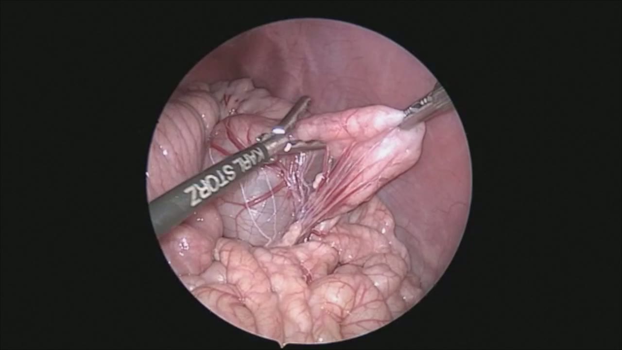
sealer demo
Dr. Steve Rothenberg · 3 min · Published May 2026
Video
pyloric knife
Dr. Steve Rothenberg · Published May 2026
Video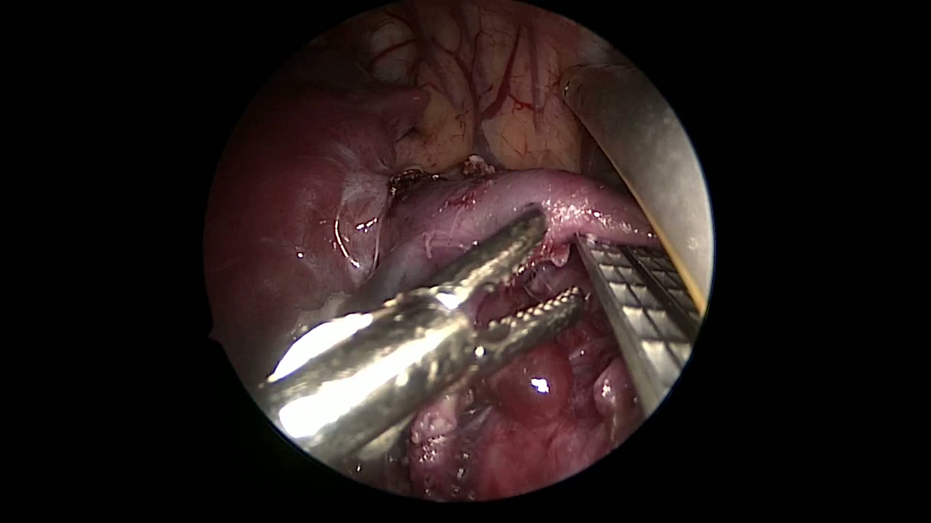
pulm artery LLL
Dr. Steve Rothenberg · 9 s · Published May 2026
Video
plication narrated
Dr. Steve Rothenberg · 6 min · Published May 2026
Video
percuvance redo nissen
Dr. Steve Rothenberg · 5 min · Published May 2026
Video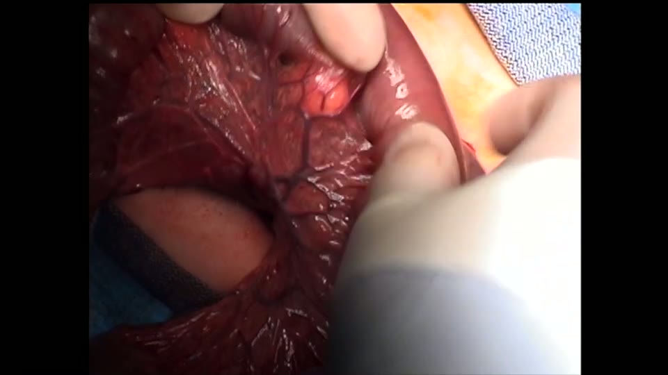
open bowel anast
Dr. Steve Rothenberg · Published May 2026
Video
Pediatric Surgical Oncology Research Collaborative (PSORC): Studying Rare Pediatric Tumors
56 s · Published May 2026
Video
Update Course Rewind 2025: Hirschsprung + ARM: Rare but Real
1 min · Published May 2026
Video
Update Course Rewind 2025: Hirschsprung + ARM: Rare but Real
1 min · Published May 2026
Video
Pooling Patients to Study Rare Pediatric Tumors: An Introduction to PSORC
56 s · Published May 2026
Video
The fetal frontier: A review of current and emerging fetal therapies for genetic diseases
44 s · Published May 2026
Video
Indocyanine green assists with sentinel lymph node mapping in pediatric and adolescent patients
1 min · Published May 2026
What the experts said
Airport X-ray scanners deliver 11 nanosieverts of radiation, which is 11 billionths of a sievert.
Background radiation from radon and cosmic sources is approximately 3 millisieverts per year.
A typical pediatric chest CT delivers 1 to 10 millisieverts, often 1-3 mSv, comparable to one year of background radiation.
CT radiation exposure in pediatric institutions has decreased dramatically with new image-gently criteria and technology improvements.
Data on childhood malignancy risk from childhood CT scans (not atomic bomb data) increased one speaker's concern about CT radiation.
Some pediatric institutions now perform sinus CT at doses lower than conventional sinus radiographs.
Sensitivity and specificity of inspiration-expiration films for airway foreign bodies are imperfect.
Physical examination does not reliably exclude airway foreign body; even normal exam does not rule it out.
A foreign body of any significance would be very unusual to have absolutely no findings on physical exam.
Volumetric chest CT for foreign body can be performed without sedation or IV contrast, and motion artifact does not prevent foreign body visualization.
At Mike's institution, 12-15 patients have undergone volumetric CT for suspected foreign body with 100% concordance between CT and bronchoscopy when bronchoscopy was performed.
CT can show air trapping distal to a foreign body and pinpoint the exact bronchial location, aiding bronchoscopic planning.
A chest radiograph delivers approximately 0.02 millisieverts; 50 chest X-rays equal one millisievert.
Performing bronchoscopy in a child with respiratory symptoms (who may have bronchiolitis or asthma rather than foreign body) carries higher anesthetic risk.
CT can visualize vegetable matter, peanuts, and most radiolucent foreign bodies because they opacify the air column; only objects with the same density as air throughout would be invisible.
Families and consultants frequently express concern about CT radiation and anesthesia risk, requiring repeated explanations even when physicians believe risks are low.
If an intussusceptum shows no movement during the first reduction attempt (especially if located in sigmoid colon or splenic flexure), second attempts are usually unsuccessful.
If the intussusceptum moves to the ileocecal valve on first attempt, second attempts are usually successful.
Delay between first and second reduction attempts ranges from 15 minutes to overnight across institutions, with most around 1-3 hours; success rates are approximately 50% regardless of delay duration.
Filling the abdomen of a trauma patient with oral contrast before potential surgery is undesirable.
Fluid-filled bowel loops serve as a natural contrast agent on CT, making oral contrast unnecessary in most cases.
Oral contrast administration delays CT scanning: contrast must be ordered from pharmacy, patients may refuse or vomit it, and NG tube placement may be required, turning a 30-minute scan into a 6-hour process.
In partial small bowel obstruction, proximal loops are slightly more dilated than distal loops and contain fluid, allowing identification of transition zones without oral contrast.
Oral contrast may be helpful in children under age one due to minimal mesenteric fat, and for MR enterography in Crohn disease.
MRI costs approximately double to triple the cost of CT.
Children age 6 and under typically require sedation for MRI; age 7 and over typically do not.
Ultrafast MRI sequences for appendicitis can be completed in 6 minutes, but table preparation time adds significantly to total scan time.
CT can identify closed-loop obstruction, mesenteric vessel swirling (suggesting volvulus), and bowel wall enhancement abnormalities that change acute management from observation to immediate surgery.
In one case, CT identified a closed loop with dead bowel in a patient initially scheduled for upper GI/small bowel follow-through, leading to OR within 30 minutes.
The 2011 American Association of Physicists in Medicine position statement concluded that risks of medical imaging at 50-100 millisieverts are low and may be non-existent.
AAPM stated that predictions of cancer risk from low-dose imaging in the media are not scientifically supported and may cause patients to forgo beneficial CT scans.
In one study, 30-50% of patients with confirmed airway foreign bodies had normal chest radiographs.
Decubitus views for foreign body detection did not improve sensitivity and decreased specificity compared to standard radiographs.
Expiration views improved sensitivity for foreign body detection but did not improve specificity.
In a 2004 study of 23 patients, low-dose CT detected all 15 foreign bodies confirmed at bronchoscopy with no discordance.
Most institutions perform delayed second attempts at intussusception reduction after initial failure, unless peritoneal signs or perforation are suspected.
Good trauma studies show oral contrast is not needed for abdominal CT in trauma.
Oral contrast is not used for trauma or appendicitis at most institutions; selective use includes partial small bowel obstruction.
Two papers and a Cochrane review suggested oral contrast may be therapeutic for adhesive small bowel obstruction, but this was based on small bowel follow-through studies, not CT, and is not established in pediatric literature.