


From
StayCurrentMD
Laryngotracheal Stenosis
With Dr. Michael Rutter · hosted by Dr. Brittany Levy
Part of
Aerodigestive / ENT 27 items
Chapter 1 of 8 · Fundamentals
Historical evolution
Historical evolution of airway stenosis management
Expert statements on this page
No expert statements were drawn from this page.
Host summaries · secondary, not cited in answers
Educational content from recorded physician discussions — not medical advice. Talk to your (or your child's) care team about your situation.
Video
Surgeon annual volume impacts recurrence rates of pediatric inguinal hernia repairs: A multi-institutional study
54 s · Published Feb 2026
Podcast
Journal of Pediatric Surgery Article Review: 2nd Quarter (Apr-Jun) 2025
16 min · Published Aug 2025
Video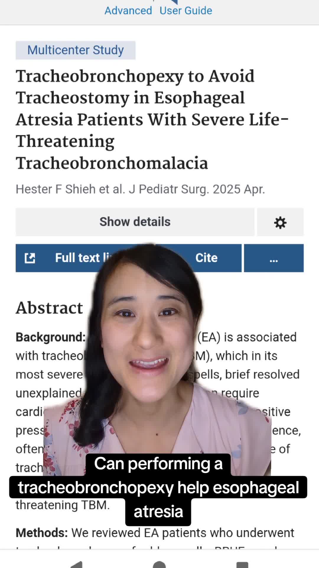
Tracheobronchopexy to Avoid Tracheostomy in Esophageal Atresia Patients With Severe Life-Threatening Tracheobronchomalacia
50 s · Published Apr 2025
Video
QUAD #27 - Multidisciplinary - How Do Teams Enhance Outcomes by the CCHMC ADEC Team
CCHMC Pediatric Surgery · 10 min · Published Apr 2025
Video
QUAD #25: Medical Management of Minor Laryngeal Clefts with Dr. Greg Burg
CCHMC Pediatric Surgery · 7 min · Published Feb 2025
Video
QUAD #22: What is CHARGE syndrome? with Dr. Catherine Hart
CCHMC Pediatric Surgery · 59 s · Published Dec 2024
Video
Intestinal Atresia Types Explained: Grossfeld Classification for Pediatric Surgery
1 min · Published Sep 2026
Video
Derivation and validation of the Pediatric Community-Acquired Pneumonia Severity (PedCAPS) score: A prospective cohort study
50 s · Published Sep 2026
Video
FETO for Late-Diagnosed Severe Congenital Diaphragmatic Hernia (CDH) at Cincinnati Children's with Dr. Beth Rymeski
4 min · Published Sep 2026
Video
Severe Congenital Diaphragmatic Hernia (CDH) Case: FETO Management & Unexpected Findings with Dr. Beth Rymeski
3 min · Published Sep 2026
Video
Fetoscopic Endoluminal Tracheal Occlusion (FETO) Procedure: Step-by-Step Guide with Dr. Beth Rymeski
4 min · Published Sep 2026
Video
Choledochal Cyst Types Explained: Pediatric Surgery Fundamentals
1 min · Published Sep 2026
What the experts said
Prior to the 1970s, bougie dilation using progressively larger dilators was the mainstay of airway stenosis management but caused significant shear forces and mucosal damage.
The development of open airway surgery in the 1970s allowed for costocartilage grafts and laryngotracheal reconstruction.
In the 21st century, endoscopic airway surgery is experiencing a resurgence and often complements open surgery.
The balloon is inflated to rated burst pressure and held for two minutes or until oxygen saturations drop to 90%, whichever happens first.
When dilating a balloon in a stenosis, pressure keeps dropping for about 90 seconds as the fibrous tissue is stretched open, requiring continuous addition of water to the syringe pump.
Kenalog injection with orotracheal injector set followed by division of scar bands in a Mercedes-Star incision with a blitzer knife is used as an adjunctive procedure.
The technique for scar division is to place the blitzer knife with the point away from the airway, get it into the stenosis, turn it 180 degrees and cut towards the lumen for a better result.
Endoscopic dilation works for stenosis at multiple levels - if you can do it in the larynx, you can do it in the trachea.
Current knowledge gaps include what size balloon to select, how much pressure is appropriate, how long to leave it inflated, when to repeat it, how often to repeat it, who should not be dilated, and when to use adjunctive procedures.
The protocol for dilation is usually three or four times at seven to ten day intervals for established scar.
On the second dilation, adjunctive procedures may include Kenalog injection, scar tissue division, increasing balloon size, or nebulizing cipridex if there are significant raw areas after dilation.
If after five dilations you are not winning, you should take a step back and think about doing something else.
The ideal candidate for balloon dilation has thin scar and young scar; thicker or established scar may require adjunctive procedures like scar division or steroid injection.
The formula for balloon sizing is to take the outer diameter of an age-appropriate endotracheal tube and add one millimeter for the larynx or two millimeters for the trachea.
A free app has been developed to help choose the right size balloon to minimize risk.
Open airway reconstruction requires an easily exposed larynx and is easier in a patient with a tracheotomy, but not required as long as the child can be kept spontaneously breathing under anesthesia.
The open procedure involves harvesting rib, exposing the larynx, placing vocal cord spreaders, dividing the posterior cricoid with a sickle knife and micro scissors, and may require dividing the inter-arytenoid muscle.
A balloon can be placed anteriorly and inflated to push the cartilage graft into place during open reconstruction.
For minor laryngeal webs, endoscopic repair is feasible and does not necessarily require a tracheotomy tube.
After dividing web scar tissue with a sickle knife, a keel must be placed to prevent raw mucosal surfaces from re-adhering.
The keel placement technique uses a keith needle threaded through silastic, taken out using a hollow angiocath as a guide.
The keel is kept in place for 10 days to allow tissue to re-mucosalize without re-adherence or risk of re-stenosis from scar tissue.
Balloon dilation should not be performed on complete tracheal rings due to rupture risk.
Balloon dilation achieves nothing in tracheomalacia and is not useful for tracheal A-frame deformity or elliptical cricoid because these are framework problems.
Endoluminal dilation is best for patients with intraluminal scar and an intact framework.
Balloon dilation delivers all dilation radially with no shear forces involved and can deliver precise high pressure dilation at low risk when the right size balloon is chosen.
Balloon dilators are single use and expensive, can be difficult to control when filling to high pressure, and are slippery with risk of watermelon seeding.
Some patients have complete and sustained improvement after a single dilation.
For patients requiring a second dilation, adjunctive scar removing procedures can be beneficial.
At Cincinnati Children's Hospital, endoscopic dilations have been performed for about 20 years with thousands of patients dilated.
In patients with intact laryngeal tracheal exoskeleton with fresher thin webs, endoscopic dilation works well, but thick and fixed scar may need adjuvant procedures or open procedure if sequential dilations are unsuccessful.
If you choose the right size balloon, the risk of balloon dilation is extraordinarily low.
Cases where balloon dilation is not enough include posterior glottic stenosis, greater than grade three posterior subglottic stenosis, and bilateral vocal cord paralysis.
Significant force is required to anchor the graft under the cut edges of the cricoid.