


From
StayCurrentMD
Laparoscopic Repair of Paraesophageal Hernia
Chapter 1 of 5 · Case-Based Learning
Case presentation
Case Presentation and Preoperative Imaging
Expert statements on this page
No expert statements were drawn from this page.
Host summaries · secondary, not cited in answers
Educational content from recorded physician discussions — not medical advice. Talk to your (or your child's) care team about your situation.
Video
Intestinal Atresia Types Explained: Grossfeld Classification for Pediatric Surgery
1 min · Published Sep 2026
Video
Derivation and validation of the Pediatric Community-Acquired Pneumonia Severity (PedCAPS) score: A prospective cohort study
50 s · Published Sep 2026
Video
FETO for Late-Diagnosed Severe Congenital Diaphragmatic Hernia (CDH) at Cincinnati Children's with Dr. Beth Rymeski
4 min · Published Sep 2026
Video
Severe Congenital Diaphragmatic Hernia (CDH) Case: FETO Management & Unexpected Findings with Dr. Beth Rymeski
3 min · Published Sep 2026
Video
Fetoscopic Endoluminal Tracheal Occlusion (FETO) Procedure: Step-by-Step Guide with Dr. Beth Rymeski
4 min · Published Sep 2026
Video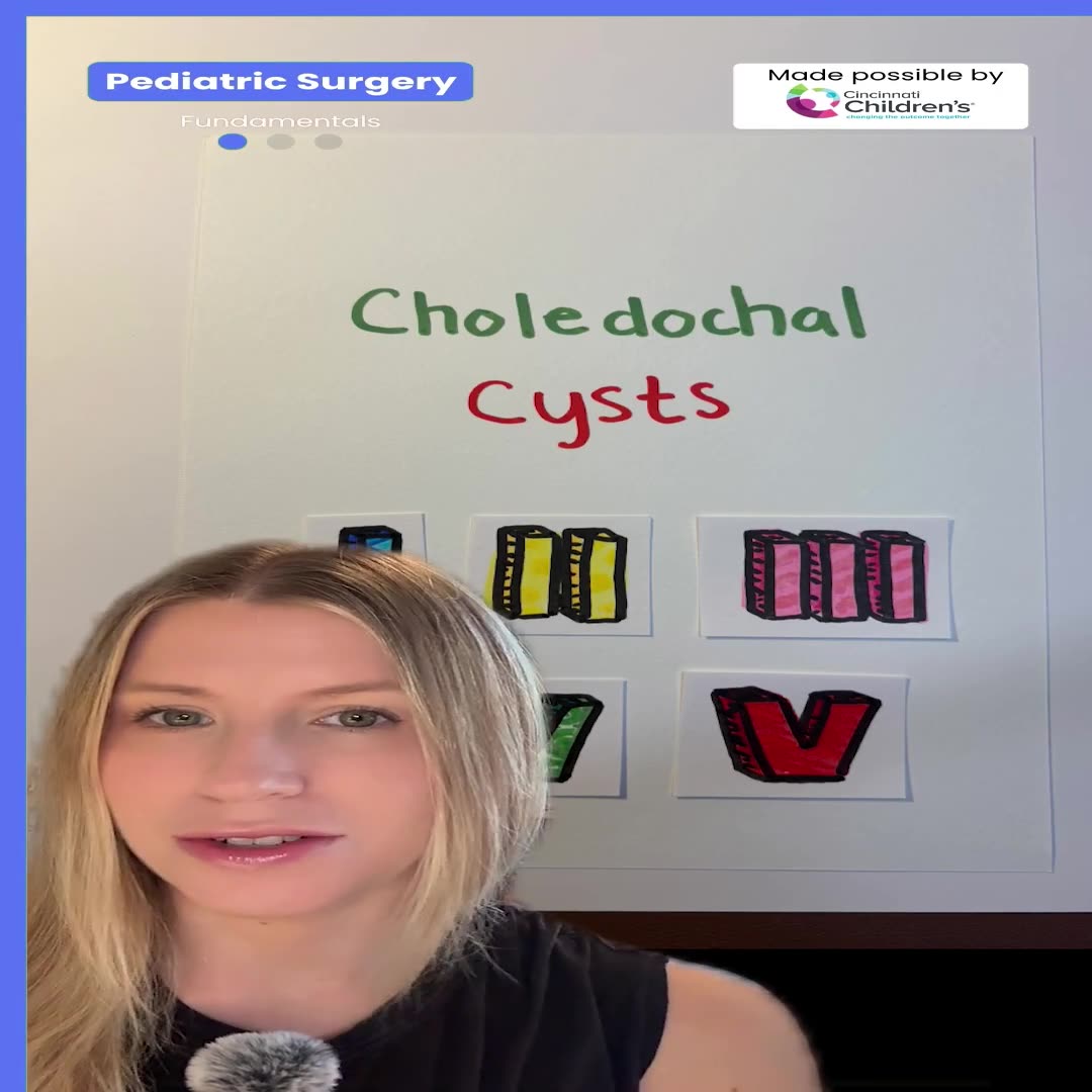
Choledochal Cyst Types Explained: Pediatric Surgery Fundamentals
1 min · Published Sep 2026
What the experts said
The most challenging part of the procedure was complete excision of the redundant adherent sac and total reduction of the distal esophagus, gastroesophageal junction, and stomach
Paraesophageal hernias are the rarest diaphragmatic hernias in children
The patient is an 18-month-old otherwise healthy girl with a type 3 hiatal hernia
The hernia was discovered on chest X-ray done for suspicion of a swallowed foreign body, revealing a right thoracic mass
The patient had poor appetite but no other GI or respiratory symptoms
CT scan revealed a large posteromedial diaphragmatic hernia containing a dilated, redundant stomach
The laparoscopic repair steps are: exposure of the defect, excision of the sac, reduction of the viscera, repair of the hernia, and fundoplication
Five 5-millimeter trocars were used with surgeon and assistant positioned at the foot of the patient
Exposure of the defect is maintained by a grasper on the diaphragm introduced through an epigastric trocar
Dissection of the sac is necessary to reduce the viscera and visualize the full defect
Sharp dissection is used to separate the sac from both the right and left diaphragmatic crura
The superior short gastric vessels are divided to completely mobilize the cardia and fundus and to aid with fundoplication
The posterior attachments of the stomach to the hiatus are divided after the stomach is freed from the spleen
Complete excision of the sac facilitates complete reduction of the GE junction and stomach
The hiatus is repaired with interrupted 2-0 braided non-absorbable suture
The anteriormost suture is placed first to minimize tension
Care is taken not to constrict the esophagus when closing the posterior defect
Sutures are placed between the esophagus and both the right and left crura to decrease the chance of recurrence
A 360-degree floppy fundoplication completes the procedure
The patient was started on oral intake the next day and discharged on the 3rd postoperative day
At 4-month follow-up, the patient was thriving with good appetite and weight had improved from the 20th to the 50th percentile