


From
StayCurrentMD
Hirschsprung Disease: Pathology Aspect
With Dr. Margaret H. Collins & Dr. Alberto Peña & Dr. Raj Kapoor & Dr. Reyes Mujica · hosted by Dr. Todd Ponsky
Chapter 1 of 7 · Diagnosis & Workup
Biopsy age limits
Suction biopsy reliability in premature infants and age limits
Expert statements on this page
No expert statements were drawn from this page.
Host summaries · secondary, not cited in answers
Educational content from recorded physician discussions — not medical advice. Talk to your (or your child's) care team about your situation.
Video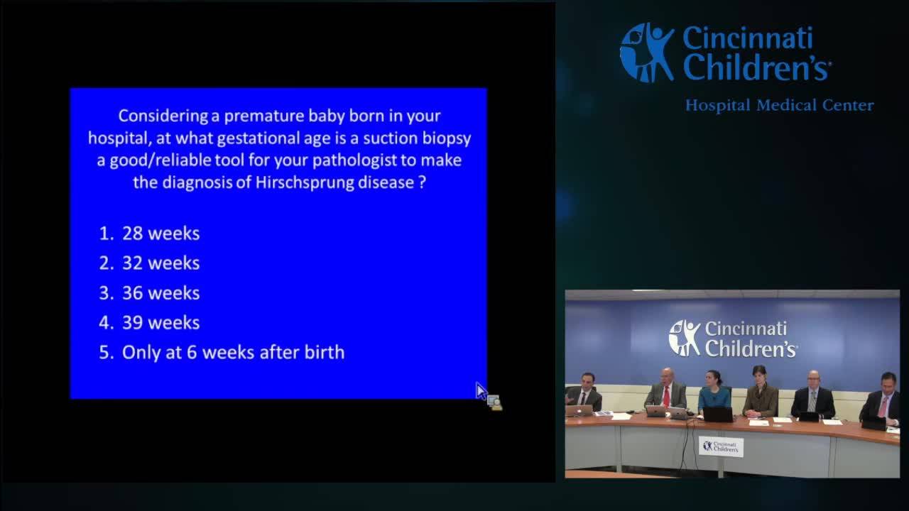
CinciHirsch - Pathology of Hirschprung Disease
44 min · Published Nov 2018
Video
Hirschsprung Disease Rapid Fire: Update Course 2015
CCHMC Pediatric Surgery · 12 min · Published Nov 2015
Video
Bowel Management for Hirschsprung's Disease Patients: Pediatric Bowel...
Dr. Todd Ponsky · 34 min · Published May 2013
Podcast
Colorectal Quiz: Episode 46
Marc Levitt · 29 min · Published Apr 2025
Video
Colorectal Quiz: Episode 46 - Hirschsprung's Disease
Marc Levitt · 29 min · Published Apr 2025
Video
Hirschsprung Disease: Update Course 2015
CCHMC Pediatric Surgery · 7 min · Published Nov 2015
Video
Validation of an anorectal malformation trainer - Can a high-fidelity model simulate real life?
1 min · Published Jun 2026
Video
Complications and Long-Term Outcomes of Patients With Cloacal Malformation After Bowel Neovagina...
1 min · Published Jun 2026
Video
Safety and utility of long-acting steroid injection for management of post-operative stricture...
1 min · Published Jun 2026
Video
Association Between Social Determinants of Health and Choice of Urinary Reconstruction in Children
1 min · Published Jun 2026
Video
The Perineal Body Preserving PSARP (PPP)
11 min · Published Jun 2026
Video
Update Course Rewind 2025: Hirschsprung + ARM: Rare but Real
1 min · Published May 2026
Video
Intestinal Atresia Types Explained: Grossfeld Classification for Pediatric Surgery
1 min · Published Sep 2026
Video
Derivation and validation of the Pediatric Community-Acquired Pneumonia Severity (PedCAPS) score: A prospective cohort study
50 s · Published Sep 2026
Video
FETO for Late-Diagnosed Severe Congenital Diaphragmatic Hernia (CDH) at Cincinnati Children's with Dr. Beth Rymeski
4 min · Published Sep 2026
Video
Severe Congenital Diaphragmatic Hernia (CDH) Case: FETO Management & Unexpected Findings with Dr. Beth Rymeski
3 min · Published Sep 2026
Video
Fetoscopic Endoluminal Tracheal Occlusion (FETO) Procedure: Step-by-Step Guide with Dr. Beth Rymeski
4 min · Published Sep 2026
Video
Choledochal Cyst Types Explained: Pediatric Surgery Fundamentals
1 min · Published Sep 2026
What the experts said
Ganglion cells are present in the rectal submucosa at 28 weeks gestation normally, though they do not necessarily look like mature ganglion cells.
An experienced pediatric pathologist will be able to recognize immature ganglion cells at 28 weeks gestation.
Suction rectal biopsies to rule out Hirschsprung's disease on 28 week gestation newborns are extremely rare.
The suction rectal biopsy is diagnostic and can be diagnostic for a patient of any age.
The failure rate for the suction rectal biopsy increases after one year of age.
Beyond infancy, there is increased separation of the ganglia as a result of the growth of the baby, contributing to suction biopsy failure.
There is increased toughness of the stroma after infancy, making it more difficult to obtain a good suction rectal biopsy.
The anal canal becomes longer and thicker after infancy, contributing to suction biopsy difficulty.
It is not a good idea to base an entire surgical procedure on the frozen section of a biopsy obtained intraoperatively.
A much better approach is to get a good full thickness rectal biopsy, not ask for a frozen section, allow the pathologist to do permanent sections and adjunctive studies if necessary, and then plan the surgical resection to begin at a future date.
When making a diagnosis of Hirschsprung disease on suction rectal biopsy, the pathologist is committing that child to losing at least some rectum.
If a suction rectal biopsy confidently shows absent ganglion cells and the surgeon finds ganglion cells at the peritoneal reflection, the patient still has short segment disease and will lose a short length of rectum.
If there is any equivocation or iffiness to the biopsy, clear communication with the surgeon is critical, and it may be necessary to rebiopsy or think of other strategies.
A lot of confident diagnosis comes back to experience: an experienced pathologist can recognize immature ganglion cells, knows how to apply and interpret ancillary studies, and knows when material is suboptimal.
Large nerves are not present in the submucosa of all cases of Hirschsprung disease, so it is possible to have a suction rectal biopsy that shows a lack of ganglion cells and does not demonstrate large nerves in the submucosa.
Total colonic aganglionosis is a classic example of Hirschsprung disease without large nerves in the submucosa.
Nerve hypertrophy may be less apparent in the very young as well as in older children.
The compulsion to do something immediately should be secondary to having a more firm diagnosis before going into the operating room.
Ancillary studies or ancillary findings are extremely helpful when you have conflicting or ambiguous results or suboptimal adequacy of a specimen.
If a patient has no ganglion cells and adequate submucosa but no hypertrophic nerves, a convincing abnormality in calretinin immunoreactivity or acetylcholinesterase staining can be enough to discount the lack of hypertrophic nerves and make the diagnosis of Hirschsprung disease.
An experienced pathologist gets a real feel for a gestalt of what are too many, too big nerves, rather than relying on nerve measurements with calipers.
The 40 micron rule (that in a young infant under 6 months of age, you shouldn't see in the distal rectum nerves greater than 40 microns in diameter) is generally true at that young age, but there are rare exceptions, and in older age kids that rule does not hold.
Complete communication between surgeon and pathologist is needed to reduce the number of difficulties and possible errors significantly.
An operation for Hirschsprung disease is too big a deal to be made on a flimsy basis.
Transition zone contains ganglion cells, but they're not in their normal distribution completely around the circumference of the bowel.
Transition zone has hypoganglionosis by definition.
Transition zone has hypertrophic nerves that can be evaluated or brought out by a glut one stain, more in the submucosa than in the myenteric plexus with or without associated ganglion cells.
A bit more controversial is submucosal hyperganglionosis with many ganglion cells in one ganglion (at least 10), in which case the submucosa can look like IND type B.
Ectopic ganglion cells can be present in what we think are normal biopsies and normally innervated bowels, so that's a bit more controversial.
A calretinin stain performed proximal to an aganglionic segment, if positive (showing nerve twigs in the lamina propria), is a sign that there are ganglion cells, even if they are not present in that particular section.
The methodology used for IND diagnosis in Europe (15 micron thick sections, at least 3 times the thickness of normal sections) has not been adopted in the United States.
The histochemical stains used for IND diagnosis are not commonly used in the United States.
There have been inconsistent diagnostic criteria for IND; the definition for the various forms of IND have changed several times over the last several decades.
The most important issue with IND is inadequate control data: one cannot look at biopsies from constipated children and conclude that any of the features are responsible for the constipation without age-matched controls from children who are not constipated.
The recommendation is that the diagnosis of IND should not be made in infants.
IND is outgrown by the age of 4.
IND is not a disorder that requires surgical therapy; it is self-correcting.
Dr. Pena could not find a single paper or study with a topographic study of neurointestinal dysplasia showing the extension of the defect.
The concept of IND is still irrelevant from a clinical standpoint, though there may be many secrets in histological characterization of motility disorders to be studied in the future.
The problem with intestinal neuronal dysplasia arose out of the need to explain the subset of patients with symptoms after Hirschsprung surgery.
IND was championed by a surgeon doing pathology, which is probably not appropriate.
IND should be put in its right place as a probably transitional period of developing in a subset of patients, but not a condition that requires surgery.
Dr. Kapoor is not as convinced as he once was that IND has been put to death, and remains open-minded as to whether there is an age-related change in the density of giant ganglia in the submucosa that is shifted in patients with Hirschsprung disease.
The best way to diagnose hypoganglionosis is to only consider the myenteric ganglion cell density, which means one has to be dealing with a resected bowel, not just a suction biopsy.
Currently, only severe hypoganglionosis is confidently diagnosed, based on long stretches of the myenteric plexus containing small ganglia (one or two ganglion cells per ganglion) with minimal neuropil.
For research purposes only, less severe forms of hypoganglionosis can be resolved with a dedicated count of ganglion cells using a particular antibody as a marker, but there is huge normal variation and a need to count large areas to get an accurate assessment, which limits the clinical value.
Multiple papers and authors have challenged if the diagnostic criteria for IND represent one end of a normal spectrum.
There is a question of whether the histopathological phenotype of IND is actually a consequence or an adaptation of downstream dysmotility rather than the cause of the dysmotility.