
Hirschsprung Disease - Imperforate Anus - Rectal Prolapse: Update Course 2015
With Dr. Jason Frischer · StayCurrentMD
Educational content from recorded physician discussions — not medical advice. Talk to your (or your child's) care team about your situation.
Video
Hirschsprung Disease Rapid Fire: Update Course 2015
CCHMC Pediatric Surgery · 12 min · Published Nov 2015
Video
Hirschsprung Disease: Update Course 2015
CCHMC Pediatric Surgery · 7 min · Published Nov 2015
Video
Hirschsprung Disease: Update Course 2013
38 min · Published Sep 2013
Podcast
Hirschsprung Disease â PediaCast 287
Marc Levitt · 38 min · Published May 2014
Podcast
Update Course Rewind: Pediatric Colorectal Consortium 2021
14 min · Published Apr 2022
Video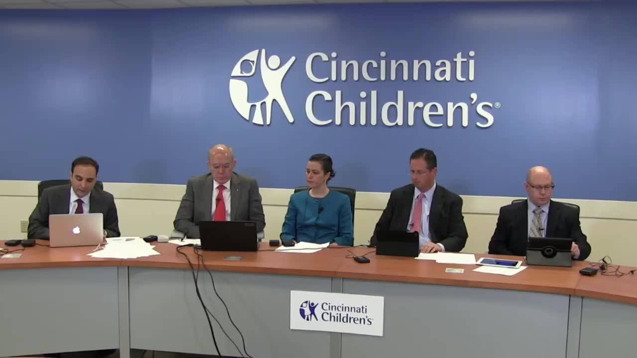
Hirschsprung Disease: Cases and Complications
103 min · Published Feb 2015
Podcast
Colorectal Quiz: Episode 47
Marc Levitt · 22 min · Published May 2025
Podcast
Colorectal Quiz: Episode 47
Marc Levitt · 22 min · Published May 2025
Podcast
Colorectal Quiz: Episode 46
Marc Levitt · 29 min · Published Apr 2025
Podcast
Colorectal Quiz: Episode 46
Marc Levitt · 29 min · Published Apr 2025
Podcast
Colorectal Quiz: Episode 43
Marc Levitt · 23 min · Published Jan 2025
Podcast
Colorectal Quiz: Episode 43
Marc Levitt · 23 min · Published Jan 2025
Video
Pediatric Surgical Oncology Research Collaborative (PSORC): Studying Rare Pediatric Tumors
56 s · Published May 2026
Video
Update Course Rewind 2025: Hirschsprung + ARM: Rare but Real
1 min · Published May 2026
Video
Update Course Rewind 2025: Hirschsprung + ARM: Rare but Real
1 min · Published May 2026
Video
Pooling Patients to Study Rare Pediatric Tumors: An Introduction to PSORC
56 s · Published May 2026
Video
The fetal frontier: A review of current and emerging fetal therapies for genetic diseases
44 s · Published May 2026
Video
Indocyanine green assists with sentinel lymph node mapping in pediatric and adolescent patients
1 min · Published May 2026
What the experts said
For Hirschsprung disease with high transition zone, laparoscopic biopsy and mobilization followed by transanal technique is the preferred approach to avoid being surprised by total colonic aganglionosis during a primary transanal procedure.
An average pediatric surgeon doing primary transanal Hirschsprung pull-through will encounter a higher-than-expected transition zone or total colonic aganglionosis approximately 1 in 10 to 1 in 15 times during their career.
A pure transanal Hirschsprung dissection is not necessarily less invasive than laparoscopic approach; prolonged torquing in the anal canal may be more traumatic than three small abdominal incisions.
For standard rectosigmoid Hirschsprung disease (6-10 cm up), transanal dissection can be completed in the same timeframe as laparoscopic mobilization if the surgeon knows the level from a good contrast study.
Post-Hirschsprung complications divide into two categories: obstructive symptoms (enterocolitis, failure to thrive, distention) and soiling issues (true incontinence vs pseudo-incontinence from constipation).
Workup for post-Hirschsprung complications includes water-soluble contrast enema and exam under anesthesia looking for dentate line position, stricture, stretched sphincter, and twists; if no anatomic cause is found, perform biopsy.
For Hirschsprung pull-through, dissection should start approximately 1 cm above the dentate line (defined as the transition from squamous to columnar epithelium, at the bottom of the anal columns).
Going only 0.5 cm above the dentate line in a newborn may result in 2.5-3 cm of aganglionic segment remaining when the child reaches 7 years old, creating an ultra-short segment Hirschsprung situation with constipation.
Injury to the dentate line during Hirschsprung pull-through can cause devastating fecal incontinence; it is better to risk leaving ultra-short segment aganglionosis (manageable with laxatives) than to injure the dentate line.
The top of the anal columns serves as a consistent anatomic landmark for Hirschsprung dissection level; measuring from anoderm to top of columns in newborns yields a reproducible distance.
In ulcerative colitis or FAP patients undergoing J-pouch creation, dissection goes right at the top of the columns, or even slightly lower if polyps are present in that region.
The dentate line is variably defined in anatomic literature and textbooks; different sources point to different locations within the anal columns when labeling the dentate or pectinate line.
Deliberately leaving a zone of aganglionosis above the dentate line is preferable to risking fecal incontinence; the resulting ultra-short segment can be overcome with laxatives, but incontinence cannot be overcome.
For newborn females with vestibular fistula and normal sacrum/renal ultrasound, initial management with dilations is appropriate; many surgeons start with this approach.
Anorectal malformations that appear widely displaced from normal anal position in the newborn period may show less displacement after a few weeks to months of growth, making initial observation reasonable.
Primary anoplasty repair in the newborn period is technically challenging; the dissection between vagina and rectum is thinner and it is harder to identify the center of the sphincter in a 2 kg baby compared to an 8-9 kg baby.
Prolonged dilations for vestibular fistula cause scarring and inflammation; dilations should be limited to maximum Hegar size 7-8 with stool softeners, as dilating to 11-12 makes subsequent repair difficult.
The traditional teaching that newborn meconium is sterile and therefore anorectal malformation repair should be done in the newborn period (before dilations introduce bacteria) is questioned; some surgeons repair at any age with stool present and feed early postoperatively with similar complication rates.
Conservative postoperative management of anorectal malformation repair includes keeping the patient NPO for approximately one week with hyperalimentation (a 'medical colostomy'), though there is no data supporting this practice over early feeding.
When vestibular fistula is discovered intraoperatively to have an absent vagina, management depends on prognosis for bowel control: if good prognosis, use graft (colon or small bowel) to create vagina and bring rectum down; if poor prognosis (sacral agenesis, tethered cord), use the rectum/fistula as vagina and bring proximal colon down as pull-through.
Rectal prolapse workup should distinguish full-thickness prolapse from partial-thickness (mucosal) prolapse based on physical examination appearance.
Testing for cystic fibrosis in children presenting with rectal prolapse is traditional teaching and a board exam answer, but in clinical practice rarely identifies new CF cases; most CF patients with prolapse are already diagnosed.
Initial management of rectal prolapse includes treating constipation and limiting toilet sitting time to 5 minutes; surgical intervention is considered after 6 months to 2 years of failed medical management.
Sclerosing agent injection for rectal prolapse has high recurrence rates; patients who fail this approach and require subsequent rectopexy have difficult reoperative dissection.
Ventral mesh rectopexy is a newer procedure popularized by the Cleveland Clinic for rectal prolapse; it involves placing mesh on the anterior rectum surface, elevating the rectum without posterior dissection, and tacking mesh to the sacral promontory.
Mesh erosion is a known complication of ventral mesh rectopexy, but the procedure can be effective for refractory rectal prolapse after multiple failed transabdominal repairs.
Ventral mesh rectopexy aims to change the angle between rectum and anal canal; younger patients with rectal prolapse have a straighter rectum-to-anal-canal angle that becomes more acute with age.
Transabdominal approaches for rectal prolapse (rectopexy with or without resection) have approximately 5% recurrence rate, compared to 15-20% recurrence for transanal approaches (transanal pull-through or Altmeier procedure).
Resection and rectopexy for rectal prolapse may be preferable when constipation control is difficult, but has higher complication rate compared to rectopexy alone.
Laparoscopic rectopexy for rectal prolapse can be performed as same-day surgery or with next-day discharge in current practice.