29 views 0 likes

Live Event Content

GCMD Space · View profile →

Gastroschisis - Clinical Practice Updates

Video Published 2020-09-14 Updated 2026-07-29

Timestops (10)

Topic Overview

A surgical discussion covering management of giant omphaloceles and large abdominal wall defects. The speakers review a two-center retrospective study of 97 survivors showing that giant omphaloceles (≥5 cm or liver-containing) carry increased risk of pulmonary hypertension, chromosomal anomalies, and delayed feeding compared to routine omphaloceles. A key finding is that five patients developed severe pulmonary hypertension after initial normal echocardiograms, all triggered by sepsis episodes. The discussion then shifts to closure techniques, including a novel sac-preserving active reduction method using hydrocolloid dressings that achieved 92% closure within 15 days in nearly 40 patients. The speakers debate whether to perform Ladd procedures during definitive closure for omphaloceles, given higher midgut volvulus risk compared to gastroschisis. A complex case of a ruptured giant omphalocele with complete liver herniation is presented, with management options ranging from biologic mesh scaffolding to eventual component separation technique for definitive closure.

Key Takeaways

  • Screen for delayed pulmonary hypertension during sepsis in giant omphalocele—5 patients developed severe PH after normal initial echo. (0:00)
  • Sac-preserving hydrocolloid technique achieved 92% closure within 15 days in nearly 40 giant omphalocele patients. (5:00)
  • Apply hydrocolloid dressing within 24 hours before sac stiffens; compressions can start at 48 hours if stable. (6:47)
  • Omphalocele carries higher midgut volvulus risk than gastroschisis due to malrotation; consider Ladd if exposing intestines. (9:16)
  • Component separation achieved fascial closure in majority of 9 children (7 days–10 years) with giant defects per UT Houston series. (26:33)

Inside this episode

Kai, the Library's AI content creator, listened to this episode and mapped who's speaking, the chapters, key claims, and cases. Every item links to the exact moment in the recording.

AI-enriched

Who's speaking

  • Sean — guest
  • Speaker 2 — host
  • Miguel Gilfoyd — guest
  • Todd — guest
  • Eric — guest
  • Saleem — guest

Chapters

  • 0:00Giant Omphalocele Physiology and Pulmonary Hypertension Risk — Review of two-center 20-year retrospective study of 97 giant omphalocele survivors, highlighting increased respiratory insufficiency, delayed feeding, and chromosomal anomalies. Discussion of delayed pulmonary hypertension triggered by sepsis in patients with initially normal echocardiograms.
  • 5:00Sac-Preserving Active Reduction Technique — Miguel Gilfoyd presents technique developed by Dr. Abello using hydrocolloid dressing to create silo without removing sac, achieving 92% closure within 15 days in nearly 40 patients. Patients kept intubated and paralyzed during serial reductions.
  • 10:31Ladd Procedure Debate for Omphalocele Closure — Discussion of whether to perform Ladd procedure during definitive omphalocele closure, based on data showing higher midgut volvulus risk in omphalocele versus gastroschisis. Debate centers on non-rotation versus malrotation terminology and clinical significance.
  • 15:48Ruptured Giant Omphalocele Case Presentation — Case of 36-week male with massive abdominal wall defect and complete liver herniation. Discussion of initial management with 10 cm silo, use of biologic mesh as scaffold, skin grafting, and tissue expander placement for eventual coverage.
  • 23:21Component Separation for Definitive Closure — Management of resulting giant ventral hernia at age 4 years. Discussion of component separation technique as alternative to permanent mesh, with review of UT Houston series showing successful closure in 9 patients aged 7 days to 10 years.

Key claims

  • 0:00Giant omphaloceles (5 cm or greater or liver in sac) have greater time to full feeds and require more TPN compared to routine omphaloceles — Sean
  • 0:00Giant omphaloceles have greater risk of respiratory insufficiency and higher incidence of chromosomal anomalies — Sean
  • 0:0056 of 97 giant omphalocele survivors were identified as having pulmonary hypertension, most diagnosed within first week of life — Sean
  • 0:00Five patients had no signs of pulmonary hypertension on first echo within first seven days but subsequently developed severe pulmonary hypertension, all associated with sepsis episodes — Sean
  • 0:00Two of the five patients with delayed pulmonary hypertension died, and one required pulmonary vasodilator for more than a year — Sean
  • 0:00One patient developed severe pulmonary hypertension 52 days after initial echo showed no pulmonary hypertension, triggered by single episode of sepsis — Sean
  • 5:00The sac-preserving hydrocolloid technique developed by Dr. Abello has been used in almost 40 patients over three years — Miguel Gilfoyd
  • 5:0097% of patients treated with sac-preserving technique achieved closure within 30 days, and 92% within 15 days — Miguel Gilfoyd
  • 5:00All patients treated with sac-preserving technique are kept in ICU, ventilated and completely paralyzed during reduction — Miguel Gilfoyd
  • 6:47The hydrocolloid dressing should be applied within first 24 hours before the sac becomes very stiff — Miguel Gilfoyd
  • 7:37Three patients with ruptured omphalocele sacs were sutured and then had hydrocolloid dressing applied successfully — Miguel Gilfoyd
  • 8:01Compressions can usually start within 48 hours if baby is stable — Miguel Gilfoyd
  • 9:16Risk of adhesive bowel obstruction is higher with gastroschisis, but risk of midgut volvulus is higher in patients with omphalocele — Sean
  • 9:16Patients with omphalocele have non-rotation or malrotation and will not have the same adhesions as gastroschisis — Sean
  • 10:31If closure technique involves exposing the intestines, Ladd procedure may be worthwhile; if sac is maintained, not worth going through sac — Sean
  • 13:10In diaphragmatic hernia repair, key move is to unroll bowel like a scroll to increase distance between ends of mesentery and decrease volvulus risk — Todd
  • 14:33Non-rotation does not exclude possibility of unfavorable anatomy with narrow base of mesentery and two ends close together — Sean
  • 16:42In gastroschisis, entire liver being out is not expected; when almost no abdominal domain exists, management becomes very challenging — Saleem
  • 20:24Spring-loaded tech silo on giant abdominal wall defect can make defect much bigger over time as forces go outward — Todd
  • 21:23Biologic mesh can be used as scaffold to allow skin epithelialization in giant defects — Saleem
  • 24:18Prolene mesh can stay in place for months without removal, with 80% of giant defects closable within 2-3 months — Miguel Gilfoyd
  • 25:37Tissue expanders placed subcutaneously can create redundant healthy skin, particularly important for cases initially treated with paint and wait — Eric
  • 26:33Component separation technique involves separating tissue at external oblique about 1 cm beyond rectus sheath on both sides, creating significant space — Sean
  • 26:33UT Houston group reported component separation use in 9 children aged 7 days to 10 years, achieving fascial closure in vast majority — Sean

Cases discussed

  • 15:4836-week gestational age male with prenatal diagnosis of abdominal wall defect, unclear if omphalocele or gastroschisis. Born with massive defect, almost no anterior abdomen, with entire liver, large amount of intestine, and stomach herniated with no visible amnion covering.

Points of disagreement

  • 10:31Whether to perform Ladd procedure during omphalocele closure
    • Sean: If closure involves exposing intestines, Ladd procedure worthwhile due to higher midgut volvulus risk in omphalocele
    • Todd: Never does Ladd in these patients; questions comparing bowel obstruction to midgut volvulus since one is lethal and one is not
  • 7:37Timing of hydrocolloid dressing application
    • Miguel Gilfoyd: Should apply within first 24 hours before sac gets stiff
    • Eric: Sometimes waits a few days until sac is no longer paper thin and see-through, still works
  • 25:09Tissue expander placement location for giant defects
    • Sean: Plastic surgeons felt intra-abdominal expanders would push contents up and out without domain
    • Eric: Uses tissue expanders primarily subcutaneously to create redundant healthy skin

Open questions

  • What is the optimal timing for tissue expander placement in giant abdominal wall defects?
  • Should intra-abdominal versus subcutaneous tissue expander placement be used for creating domain?
  • At what age is component separation technique most effective for giant ventral hernias?
  • Can the sac-preserving hydrocolloid technique be applied after waiting several days, or must it be within 24 hours?
  • What is the true incidence of midgut volvulus in non-rotated versus malrotated omphalocele patients?
  • Should all omphalocele closures that expose intestines include Ladd procedure regardless of technique?
This episode was analyzed and enriched by Kai, the Library's AI content creator. Every item links to the moment it comes from — click a timestamp to listen in context.

Massive Abdominal Wall Defect With Entire Liver Herniated: Staged Reconstruction Over Four Years

The patient case from this episode, retold from presentation to outcome with the decisions made along the way. Written by Kai from the episode transcript and reviewed before publishing.

For the care team · Case narrative · AI-written, human-reviewed

Presentation

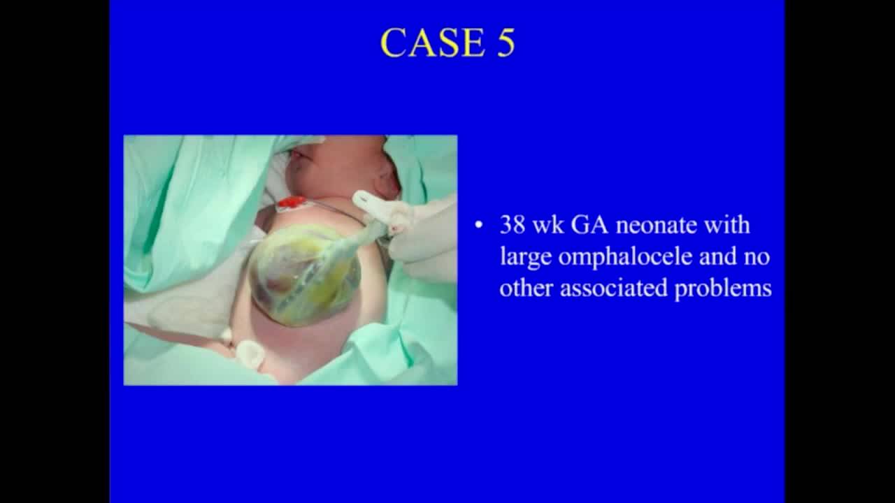
A 36-week male was born with a prenatal diagnosis of an abdominal wall defect that maternal-fetal medicine specialists could not definitively characterize as omphalocele or gastroschisis. At delivery, the infant had almost no anterior abdominal wall, with the entire liver, a large amount of intestine, the stomach, and colon herniated [case1]. No visible amnion or covering was present [case1]. The defect was located in the center of the abdomen with the umbilical cord at the upper margin [case1]. A 10-cm spring-loaded silo was placed to cover the viscera [case1].

The Decision Point

The team faced an immediate classification problem with direct management implications. In gastroschisis, the liver is not expected to be herniated 16:42. When almost no abdominal domain exists, management becomes very challenging 16:42. The spring-loaded silo presented a mechanical problem: on giant abdominal wall defects, downward pressure creates outward forces that can enlarge the defect over time 20:24. When one discussant attempted compression, "it just popped right out because the defect is so big that you can't nothing holds it in place" [q22].

The team classified this as a ruptured omphalocele and chose biologic mesh as a scaffold rather than attempting active reduction [case1]. The rationale was to allow skin epithelialization rather than risk further fascial disruption 21:23. They performed a circumcision and used that skin as an additional graft [case1]. Plastic surgeons placed tissue expanders to create redundant healthy skin, particularly important for cases initially treated with topical agents that leave poor-quality skin 25:37. The team serially resected portions of the mesh before full incorporation, gradually reducing the contents and stretching the fascia to create some abdominal domain [case1].

Outcome and Delayed Presentation

The infant survived the initial management and achieved skin coverage [case1]. At four years of age, he presented with a giant ventral hernia — his abdominal contents appeared ready to herniate with ambulation [case1]. The team performed component separation for definitive closure [case1].

The technique involves separating tissue at the external oblique approximately 1 cm beyond the rectus sheath on both sides, creating significant space 26:33. Dissection between the external oblique and the deeper muscle layers, combined with an incision on the anterior rectus sheath for an additional centimeter of mobilization bilaterally, generates substantial room for midline approximation 26:33. A group from UT Houston reported using this approach in nine children aged 7 days to 10 years, achieving fascial closure in the vast majority, though some required bridging mesh 26:33.

What the Case Changes

This case illustrates three transferable principles. First, spring-loaded silos may be mechanically unsuitable for defects with minimal abdominal domain — the outward force vector can worsen the problem you are trying to solve 20:24. Second, biologic mesh functions as a scaffold for epithelialization when primary closure is impossible, and can remain in place for months; one discussant reported 80% of giant defects closable within 2-3 months using this staged approach with prolene mesh 24:18. Third, component separation — routine in adult abdominal wall reconstruction — is applicable across the pediatric age spectrum for giant defects, including in neonates 26:33. The technique borrows from adult hernia surgery but addresses a congenital problem, and the Houston series demonstrates its feasibility even in the first week of life 26:33.

The case also demonstrates that massive abdominal wall defects with complete liver herniation occupy a gray zone between classic gastroschisis and omphalocele, and that rigid classification may matter less than recognizing when standard closure techniques will fail and staged reconstruction is required from the outset.

Takeaways from this story

  • Spring-loaded silos on giant defects create outward forces that can enlarge the defect rather than reduce it
  • Biologic mesh serves as scaffold for epithelialization; 80% of giant defects closable within 2-3 months using staged approach
  • Component separation achieves fascial closure in giant defects across all pediatric ages, including first week of life
  • Tissue expanders placed subcutaneously create redundant healthy skin, critical when initial treatment leaves poor skin quality

Keywords

Hashtags

Transcript

Comments

Loading comments…