

Combined cervical and thoracic approach for esophageal atresia with tracheopexy
With Dr. Alessandro de Alarcon · hosted by Dr. Todd Ponsky
Chapter 1 of 5
Introduction
Introduction and Conference Context
Expert statements on this page
No expert statements were drawn from this page.
Host summaries · secondary, not cited in answers
Educational content from recorded physician discussions — not medical advice. Talk to your (or your child's) care team about your situation.
Video
QUAD #2 Thoracoscopic Tracheopexy with Dr. Aaron Garrison
CCHMC Pediatric Surgery · 7 min · Published Jan 2024
Podcast
Journal of Pediatric Surgery Article Review: 4th Quarter (Oct-Dec) 2024
18 min · Published Feb 2025
Video
BOB Ped Surg 2023 - Basma Magdy, PAPSA - Presentation
Published Feb 2023
Video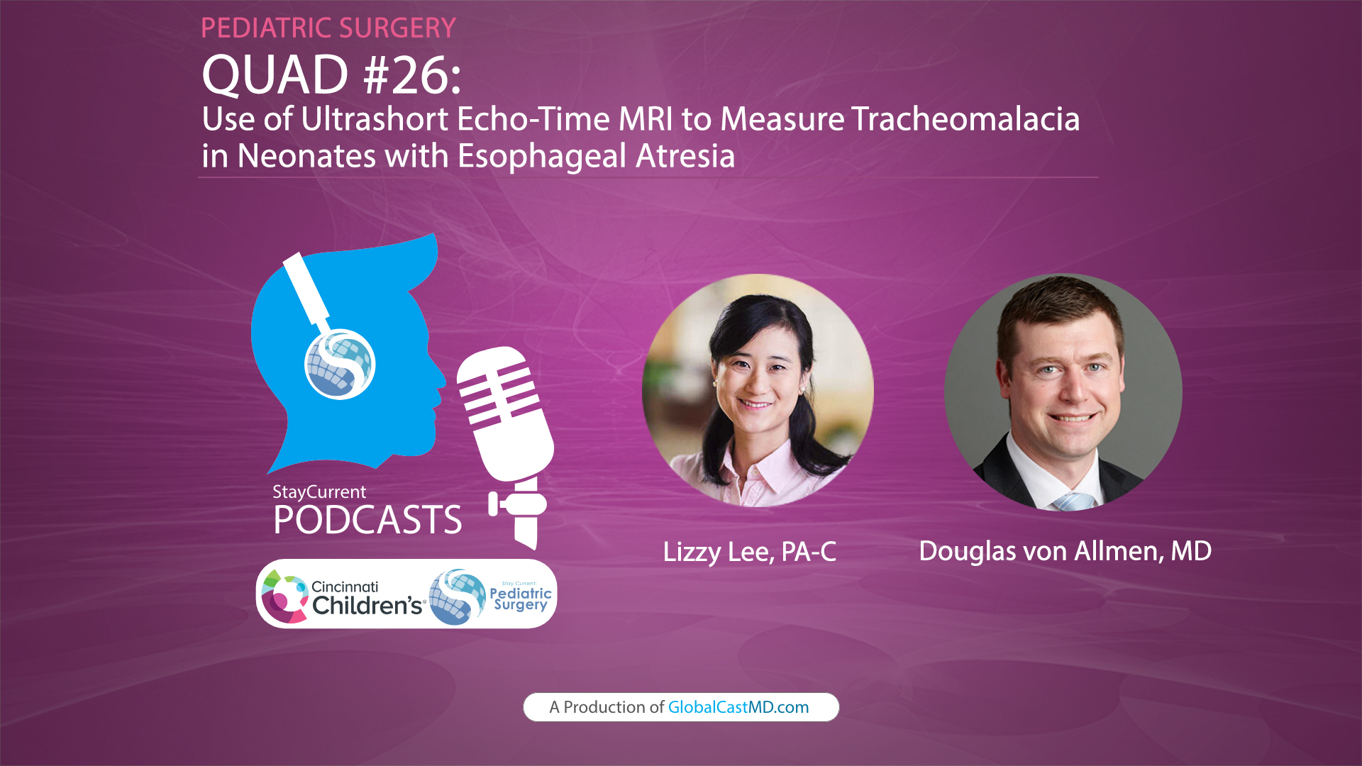
QUAD #26 - Use of Ultrashort Echo-Time MRI to Measure Tracheomalacia in Neonates with Esophageal Atresia with Dr. Douglas von Allmen
CCHMC Pediatric Surgery · 5 min · Published Feb 2025
Video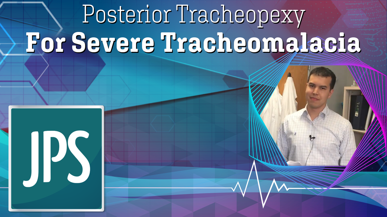
Posterior Tracheopexy For Severe Tracheomalacia
Dr. Todd Ponsky · 2 min · Published Feb 2018
Video
Esophageal Atresia/Tracheoesophageal Fistula Bronchoscopy and Surgical Technique
Dr. Todd Ponsky · 16 min · Published Nov 2021
Podcast
Preventing MRSA infections in pediatric airway surgery through preoperative screening
Alessandro de Alarcon · 8 min · Published Oct 2026
Video
QUAD #1: Cervical Tracheopexy with Dr. Alessandro de Alarcon
Dr. Todd Ponsky · 5 min · Published Dec 2023
Video
Inside Tract: Bilateral Vocal Fold Paralysis | Cincinnati Children's
Alessandro de Alarcon · Published Mar 2020
Podcast
Screening and treatment of MRSA colonization in pediatric airway surgery patients
Alessandro de Alarcon · 8 min · Published Feb 2012
What the experts said
Cincinnati Children's typically uses a combined cervical and thoracic approach for esophageal atresia cases.
Required intraoperative equipment includes a neck tray, MLB tray, and Maloney dilators versus NG tubes.
Flexible bronchoscopy during the operation is key, with the endotracheal tube placed exactly where needed for visualization.
Nasotracheal intubation is often used with the cuff positioned very high, sometimes almost at the glottis.
A NIM tube is used when possible to prevent potential injury to recurrent nerves or provide awareness when getting close to them.
The standard neck approach involves raising subplatysmal flaps and dealing with anterior compression as needed.
Aortopexy and mopexy can be added at the same time as the cervical approach.
The straps are separated to expose the airway and find the esophagus.
The Cincinnati team has learned not to be afraid of the esophagus, being able to find it, move it, and get it out of the way.
Stitches are sometimes placed in the trachea to pull it up and out of the way to visualize the posterior aspect where the spine is visible.
3-0 Prolene sutures are used, with all stitches placed before securing them down, under spontaneous ventilation conditions.
A patient who had prior thoracoscopic tracheopexy developed dysphagia from a torqued esophagus, was treated with the combined approach, achieved a good airway, and is now symptom-free.
Outcome measures include follow-up endoscopy, pulmonary function tests when patients are old enough to perform them, and imaging.
At 36 months follow-up, imaging may still show some malacia, but patients are symptomatically better.
The team is still learning what measures should be used to define good versus bad outcomes.
Managing complications including swallowing dysfunction and vocal fold paralysis is part of the care process.
Preoperative testing includes dynamic CT imaging, pulmonary function tests, microlaryngoscopy and bronchoscopy, and flexible bronchoscopy.
Residual or regrown large thymus can be removed during the cervical approach as it is in the way of the thoracoscopic approach.
In the lateral approach, the pediatric surgeon stands on the side of the airway to look for the esophagus while the ENT surgeon looks for the recurrent laryngeal nerve.
The esophagus is mobilized above the level where the surgeons aim to pexy, making it easier to place sutures exactly where needed.
A pulmonologist assists with flexible tracheal endoscopy through the endotracheal tube while stitches are being placed.
The combined approach is valuable for complicated cases or patients needing additional operations for symptom relief.
Otolaryngology involvement is important both in the procedure and during follow-up, and is easier when already built into the team.