


From
Live Event Content
Dominique Simons, MSc - Best of the Best in Pediatric Surgery 2024
With Dr. Dominique Simons
Chapter 1 of 6 · Fundamentals
Introduction
Introduction and disclosures
Expert statements on this page
No expert statements were drawn from this page.
Host summaries · secondary, not cited in answers
Educational content from recorded physician discussions — not medical advice. Talk to your (or your child's) care team about your situation.
Video
BOB Ped Surg 2023 - Bernadette Jeremiasse, IPSO - Presentation
5 min · Published Feb 2023
Video
Social Determinants of Health Influence on Survival in Wilms Tumor, Neuroblastoma, and Hepatoblastoma
1 min · Published Jan 2026
Video
Update Course Rewind: Staging and Management of Neuroblastoma 2022
Published Jun 2023
Podcast
Journal of Pediatric Surgery Article Review: The Disruption Score
12 min · Published Aug 2021
Video
The International Neuroblastoma Risk Group (INRG) staging system: an INRG Task Force report
59 s · Published Mar 2026
Podcast
The Future of Surgical Education: A King, an Ox, & Elon Musk
Todd Ponsky · 26 min · Published Oct 2026
Video
Oncology Tumor Board Showdown: Pediatric Oncology 5 Question Quiz
1 min · Published Oct 2026
Video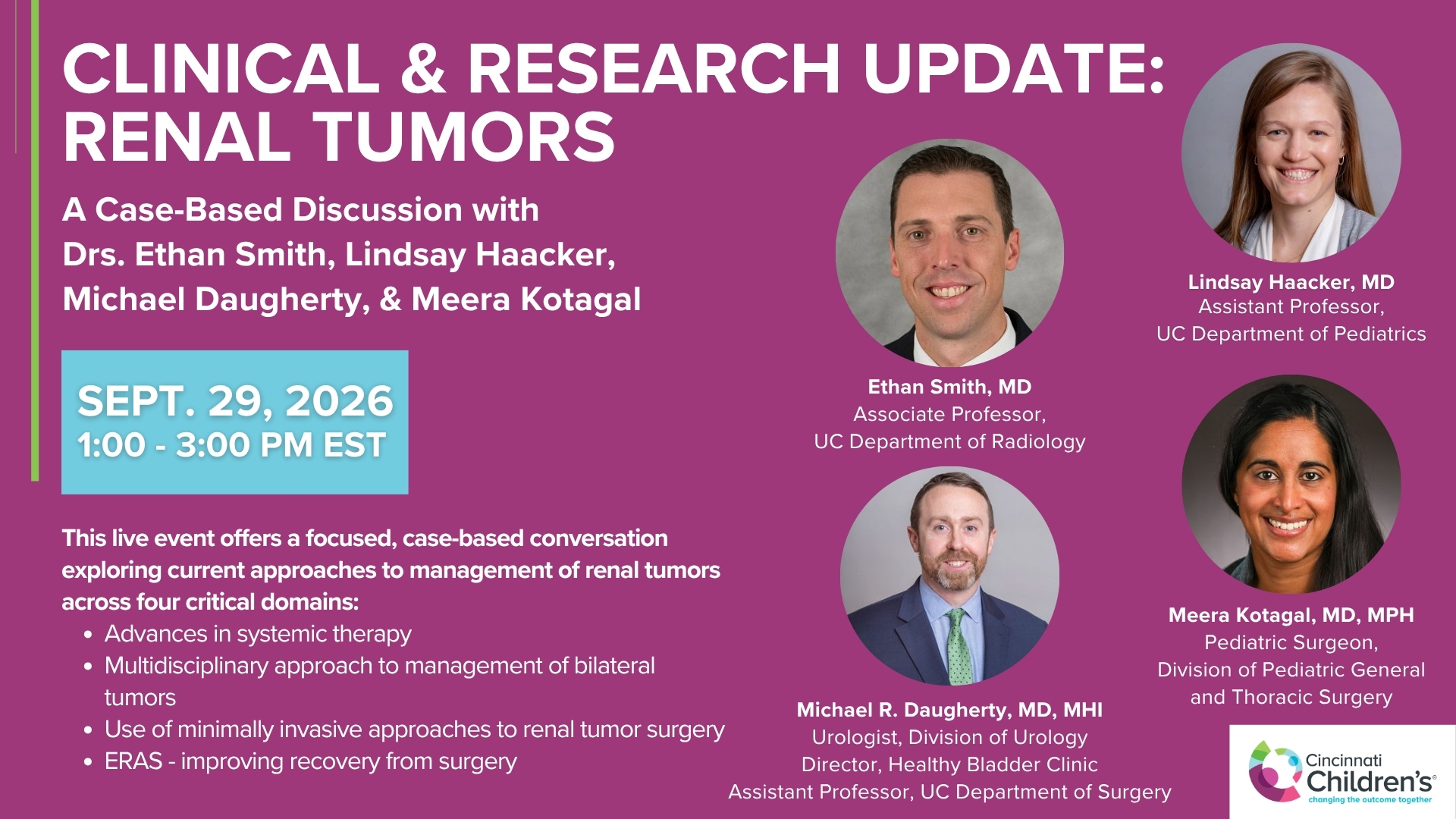
Clinical & Research Update: Renal Tumors with Drs. Ethan Smith, Lindsay Haacker, Michael Daugherty, and Meera Kotagal
71 min · Published Sep 2026
Video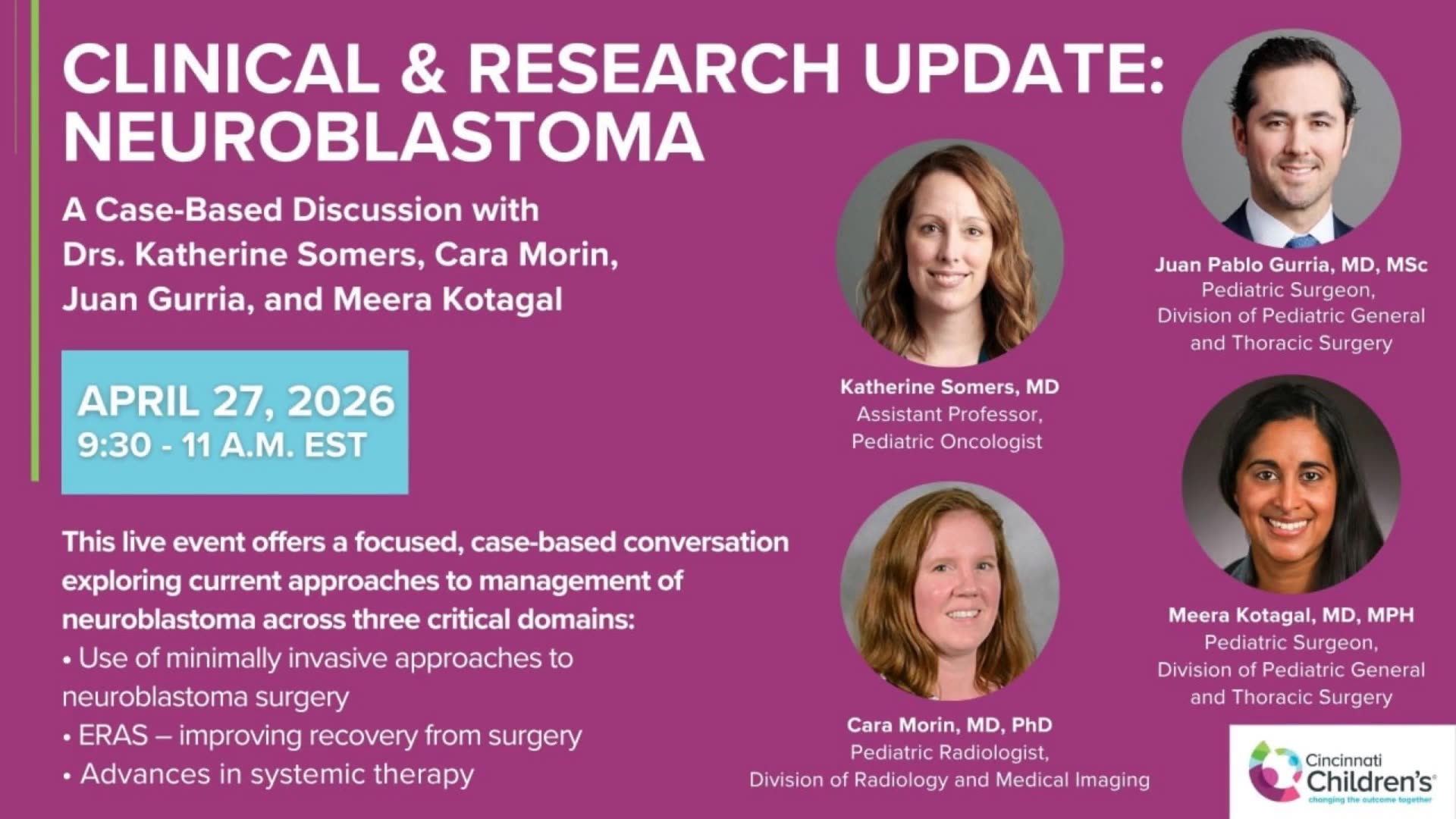
Clinical & Research Update: Neuroblastoma with Drs. Katherine Somers, Cara Morin, Juan Gurria, and Meera Kotagal
67 min · Published Sep 2026
Video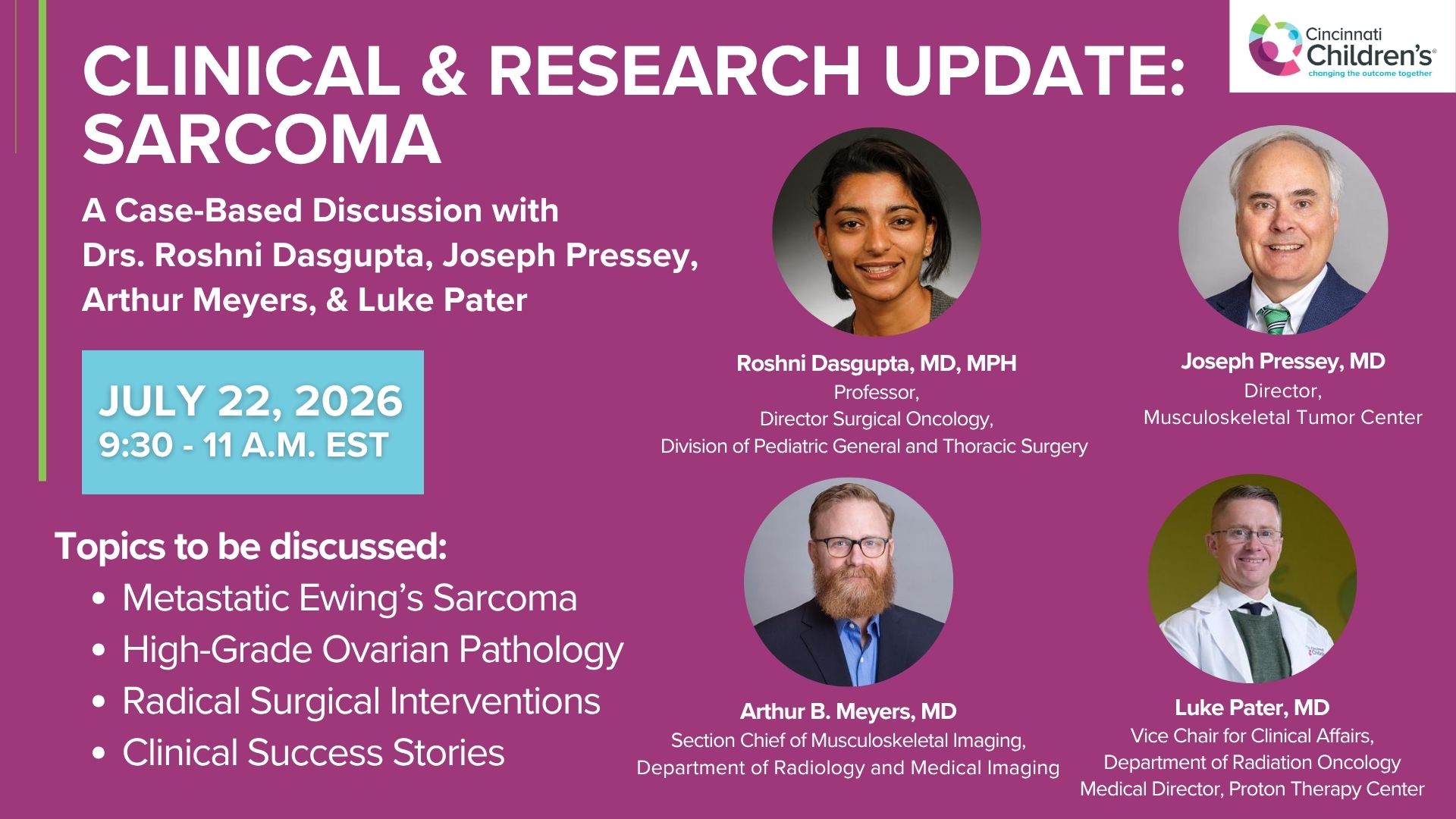
Clinical & Research Update: Sarcoma w/ Drs. Roshni Dasgupta, Joseph Pressey, Arthur Meyer, Luke Pater
66 min · Published Sep 2026
Video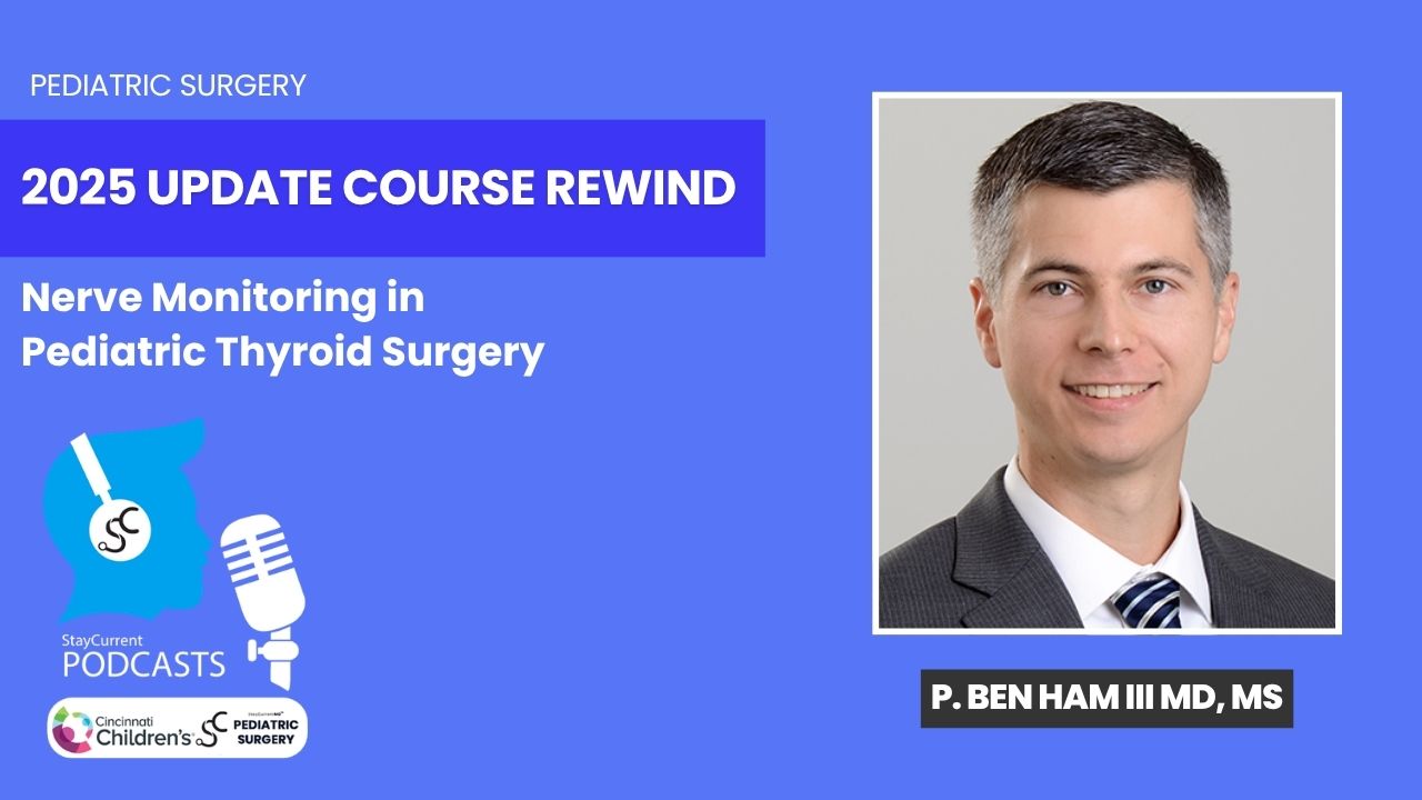
Update Course Rewind 2025: Nerve Monitoring in Pediatric Thyroid Surgery
6 min · Published Jul 2026
Video
Journal of Pediatric Surgery Article Review: June 2023, AAP Issue
Todd Ponsky · 13 min · Published Jul 2026
Only a few other public items share this expert — go deeper there →
Video
2026 Laparoscopic Pediatric Hernia Repair
123 min · Published Jun 2026
Video
Beyond the Spectrum: Diagnosis, Myths & Management - Caitlin Couch & Leslie Lopez - APP Conference 2026
50 min · Published May 2026
Video
Dysautonomia: Navigating the Journey - Martha Willis - APP Conference 2026
55 min · Published May 2026
Video
Everything You Ever Wanted to Know About G-tubes But Were Afraid to Ask - Betsy Gerrein - APP Conference 2026
30 min · Published May 2026
Video
Take My Breath Away - Carolyn Grad, Mark Washam, & Katie Holtman - APP Conference 2026
58 min · Published May 2026
Video
Central Lines...WHAT? - Richelle Guinigundo - APP Conference 2026
26 min · Published May 2026
What the experts said
Neuroblastoma is surgically challenging to resect mainly due to adhesion and encasement to important structures, heterogeneity, and therapy-induced changes.
The surgical goal for neuroblastoma is to resect more than 95% of the tumor tissue while sparing anatomical structures.
3D models of patient-specific anatomy are routinely made to prepare surgeons for neuroblastoma resection.
Complete resection of neuroblastoma is often very difficult or even impossible due to tumor location.
Surgeons face a dilemma of whether to resect more tumor tissue with possible complications such as bleeding or to leave residual tumor tissue.
Biology can be used as a biomarker for tumor vitality in neuroblastoma.
In ADC imaging, diffusion restriction is associated with viable tumor tissue and appears as a low signal.
In MIBG SPECT-CT imaging, high uptake is associated with viable tumor tissue.
MIBG uptake and diffusion restriction in neuroblastoma change over time, suggesting the biology and vitality of the tumor may also change.
ADC and MIBG scans have different 3D positions from T1-weighted MRI, requiring image registration.
Registration accuracy is evaluated using the Dice coefficient (which measures overlap between two structures) and target registration error (which measures distance between two points).
Risk group delineation uses ADC and MIBG values within the tumor volume with thresholds based on literature.
High-risk areas in the 3D model are associated with diffusion restriction and MIBG uptake.
Seven patients with abdominal neuroblastoma eligible for surgery were included prospectively.
Accurate initial registration was achieved for all patients with a median Dice of 0.81 for ADC and 0.77 for MIBG.
Median target registration error was 5.3 mm for ADC and 4.3 mm for MIBG.
Multimodal 3D models were successfully created for every patient in the study.
In some cases, ADC and MIBG risk models showed similar patterns, with the lateral side of the tumor marked as high risk and the medial side as low risk.
In other cases, ADC and MIBG identified completely different regions as high risk (e.g., cranial part high-risk on ADC versus a different region on MIBG).
The thresholds used for risk stratification are based on limited literature, which questions the reliability of the ADC and MIBG values used.
The image registration is performed using Elastix software in programming environments such as Python or MATLAB.
Segmentation of the neuroblastoma is the most time-consuming step; the actual registration takes only a few minutes.
Simmons performs the 3D model creation herself with verification from radiologists, especially in early phases.
Current practice believes that MIBG-positive areas should be resected, but discordant ADC/MIBG findings suggest this may need re-evaluation.