


Chapter 1 of 4 · Case-Based Learning
Case & positioning
Case presentation and patient positioning
Expert statements on this page
No expert statements were drawn from this page.
Host summaries · secondary, not cited in answers
Educational content from recorded physician discussions — not medical advice. Talk to your (or your child's) care team about your situation.
Video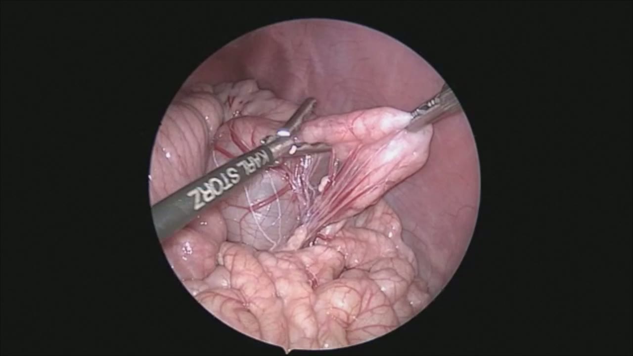
sealer demo
Dr. Steve Rothenberg · 3 min · Published May 2026
Video
pyloric knife
Dr. Steve Rothenberg · Published May 2026
Video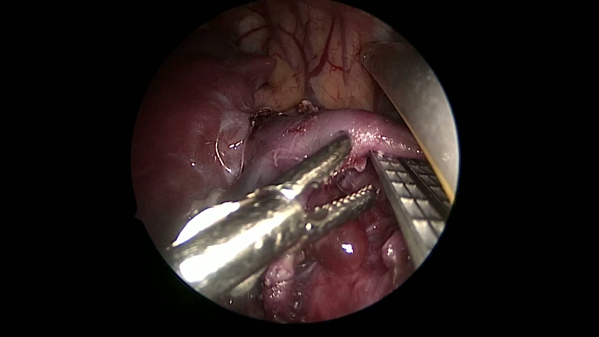
pulm artery LLL
Dr. Steve Rothenberg · 9 s · Published May 2026
Video
plication narrated
Dr. Steve Rothenberg · 6 min · Published May 2026
Video
percuvance redo nissen
Dr. Steve Rothenberg · 5 min · Published May 2026
Video
open bowel anast
Dr. Steve Rothenberg · Published May 2026
Video
Intestinal Atresia Types Explained: Grossfeld Classification for Pediatric Surgery
1 min · Published Sep 2026
Video
Derivation and validation of the Pediatric Community-Acquired Pneumonia Severity (PedCAPS) score: A prospective cohort study
50 s · Published Sep 2026
Video
FETO for Late-Diagnosed Severe Congenital Diaphragmatic Hernia (CDH) at Cincinnati Children's with Dr. Beth Rymeski
4 min · Published Sep 2026
Video
Severe Congenital Diaphragmatic Hernia (CDH) Case: FETO Management & Unexpected Findings with Dr. Beth Rymeski
3 min · Published Sep 2026
Video
Fetoscopic Endoluminal Tracheal Occlusion (FETO) Procedure: Step-by-Step Guide with Dr. Beth Rymeski
4 min · Published Sep 2026
Video
Choledochal Cyst Types Explained: Pediatric Surgery Fundamentals
1 min · Published Sep 2026
What the experts said
For thoracoscopic H-type fistula repair, the patient is placed in modified lateral decubitus position rolled prone approximately 45 degrees.
Three ports were used: one 4 mm port (later advanced to 5 mm) and two 3 mm ports.
A 4 mm 30-degree lens was used for the procedure and placed behind the tip of the scapula.
The procedure starts in the apex using a 3 mm vessel sealer for dissection and to open the pleura.
Developing a plane between esophagus and trachea below the level of the fistula is an important maneuver to help safely separate the two structures before encountering the very adherent area.
Placing a catheter across the H-type fistula can occasionally be helpful for identification, though it was not used in this case.
A 3 mm hook cautery was used to pull on fibers and separate them away from critical structures.
Dissection is carried out above the fistula so that a retro-fistula plane can be developed.
An instrument is passed around behind the fistula, taking care not to injure the esophagus or membranous portion of the trachea.
A 2-0 silk suture is passed around the fistula to help with retraction.
Previously, the technique involved dividing the fistula and then oversewing the two ends.
In this case, with the child large enough and availability of a 5 mm stapler, a decision was made to use the stapler to divide the fistula.
Care must be taken to ensure the recurrent laryngeal nerve and other important structures are not within the field of dissection.
The lower port in the posterior axillary line was enlarged to 5 mm to accommodate the stapler.
A single application of the 5 mm stapler divided the fistula, placing two rows of staples on either side.
Apical fat is taken and sewn down to cover the esophageal end of the staple line to help separate the two staple lines and prevent recurrent fistulization.
The pleura is closed over the divided fistula so that any leak would remain contained.
The lung was re-expanded and no chest tube was left.
The patient was a 3-month-old infant with multiple medical issues who had recurrent aspiration.
Upper GI study showed an H-type tracheoesophageal fistula.
There were 2 prior attempts at endoluminal ablation before this thoracoscopic repair.
The patient was examined at 2 weeks postoperatively and the fistula was assessed at 3 months.