

IPEG 2020 TOP ABSTRACT: Thoracoscopic lingulectomy for a Prenatally diagnosed CPAM in a 3 month old infant
With Dr. Steve Rothenberg
Chapter 1 of 6 · Diagnosis & Workup
Prenatal diagnosis
Prenatal diagnosis and preoperative imaging
This video is for verified healthcare professionals.Sign in to watch — the rest of this page is open.Sign in
Expert statements on this page
No expert statements were drawn from this page.
Host summaries · secondary, not cited in answers
Educational content from recorded physician discussions — not medical advice. Talk to your (or your child's) care team about your situation.
Video
Thoracoscopic Right Lower Lobectomy in a 3-Month-Old with Prenatally Diagnosed CPAM
Dr. Steve Rothenberg · 16 min · Published May 2026
Video
Clinical Symptoms Affect Treatment and Prognosis in Pediatric Patients with Congenital Pulmonary Airway Malformation
55 s · Published Dec 2023
Video
Thoracoscopic Left Lower Lobectomy for Intralobar Sequestration in a 4-Month-Old
Dr. Steve Rothenberg · 4 min · Published May 2026
Video
Should We Resect Asymptomatic CPAM Flake vs Langer
Dr. Steve Rothenberg · 14 min · Published May 2019
Video
Congenital Lung Malformations: CPAM, BPS, CLE, and Bronchogenic Cyst Explained
3 min · Published Oct 2026
Video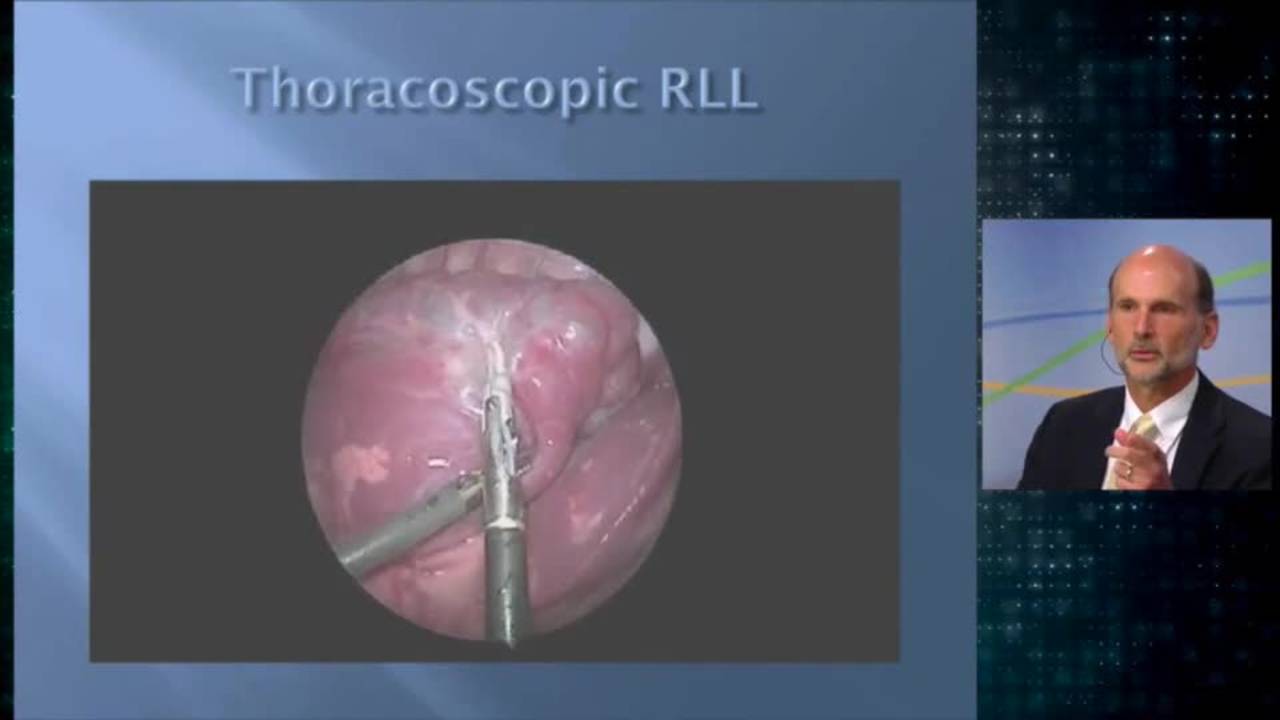
Thoracoscopic Right Lower Lobe Cystic Lesion Excision: Update Course 2014
Dr. Steve Rothenberg · 11 min · Published Sep 2014
Video
Laparoscopic Mesenteric and Short Gastric Dissection with a Vessel Sealer
Dr. Steve Rothenberg · 3 min · Published May 2026
Video
Laparoscopic Pyloromyotomy with a Reusable Handle and Disposable Blade
Dr. Steve Rothenberg · Published May 2026
Video
Thoracoscopic Stapling of the Left Lower Lobe Pulmonary Artery with a 5 mm Stapler
Dr. Steve Rothenberg · 9 s · Published May 2026
Video
Laparoscopic Plication of a Right Diaphragmatic Eventration in a 3-Month-Old
Dr. Steve Rothenberg · 6 min · Published May 2026
Video
Laparoscopic Redo Nissen Fundoplication in a 30 kg Child with the 3 mm Percuvance System
Dr. Steve Rothenberg · 5 min · Published May 2026
Video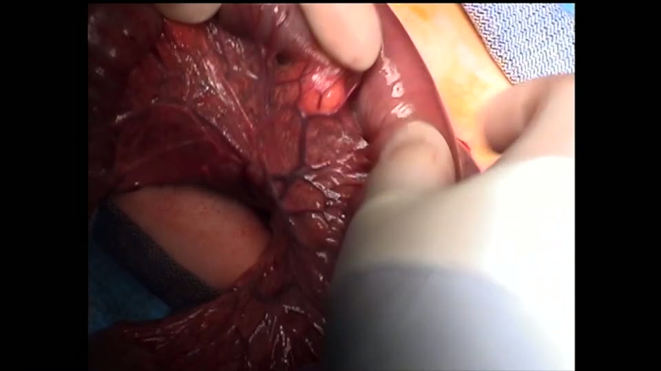
Open Bowel Anastomosis
Dr. Steve Rothenberg · Published May 2026
What the experts said
CPAM was diagnosed by prenatal ultrasound at 26 weeks
MRI at 29 weeks confirmed the CPAM diagnosis
Repeat ultrasound at 36 weeks showed an enlarging CPAM
CT scan at 3 months of age showed persistence of the CPAM in the lingula
An incomplete fissure was found upon entering the left chest
The fissure was developed using the 3 millimeter vessel sealer
The fissure can be taken in a systematic fashion with minimal blood loss and no injury to surrounding structures despite being quite incomplete
The 3 millimeter vessel sealer is used to take vessels with two separate seals placed at least 3 to 4 millimeters apart, then the vessel is divided between the seals
Partial division is accomplished first to ensure that the seals are intact, and once the lumen is seen, then the division is completed
Segmental resection of the lingula is accomplished fairly easily once all vascular and bronchial structures are taken
Hyperinflation of the lingula persists even though the rest of the lung is collapsed because of right main stem intubation
In this case, the bronchus to the lingula lies on top of the artery
The bronchus, because of its small size, is taken with a singular 5 millimeter clip
After bronchus and artery division, a line of demarcation begins to be seen between the lingula and the rest of the upper lobe
Separation between the lingula and the superior segment of the upper lobe is started with the 3 millimeter sealer and completed with the 5 millimeter stapler once the plane is clearly seen
Three applications of the 5 millimeter stapler are used to completely divide the lingula from the remnant of the upper lobe
No air leak was present after resection with lower lobe and upper lobe clearly inflating
Chest tube was removed on postoperative day one and the patient was discharged home