


From
StayCurrentMD
Laryngeal Clefts
With Dr. Michael Rutter & Dr. Alessandro Dialicon · hosted by Dr. Rod Gerardo
Chapter 1 of 6 · Fundamentals
Cleft classification
Introduction to laryngeal clefts and classification systems
Expert statements on this page
No expert statements were drawn from this page.
Host summaries · secondary, not cited in answers
Educational content from recorded physician discussions — not medical advice. Talk to your (or your child's) care team about your situation.
Video
QUAD #20: Endoscopic Repair of Minor Laryngeal Clefts with Dr. Catherine Hart
CCHMC Pediatric Surgery · 6 min · Published Nov 2024
Video
Esophageal Atresia/Tracheoesophageal Fistula Bronchoscopy and Surgical Technique
Dr. Todd Ponsky · 16 min · Published Nov 2021
Video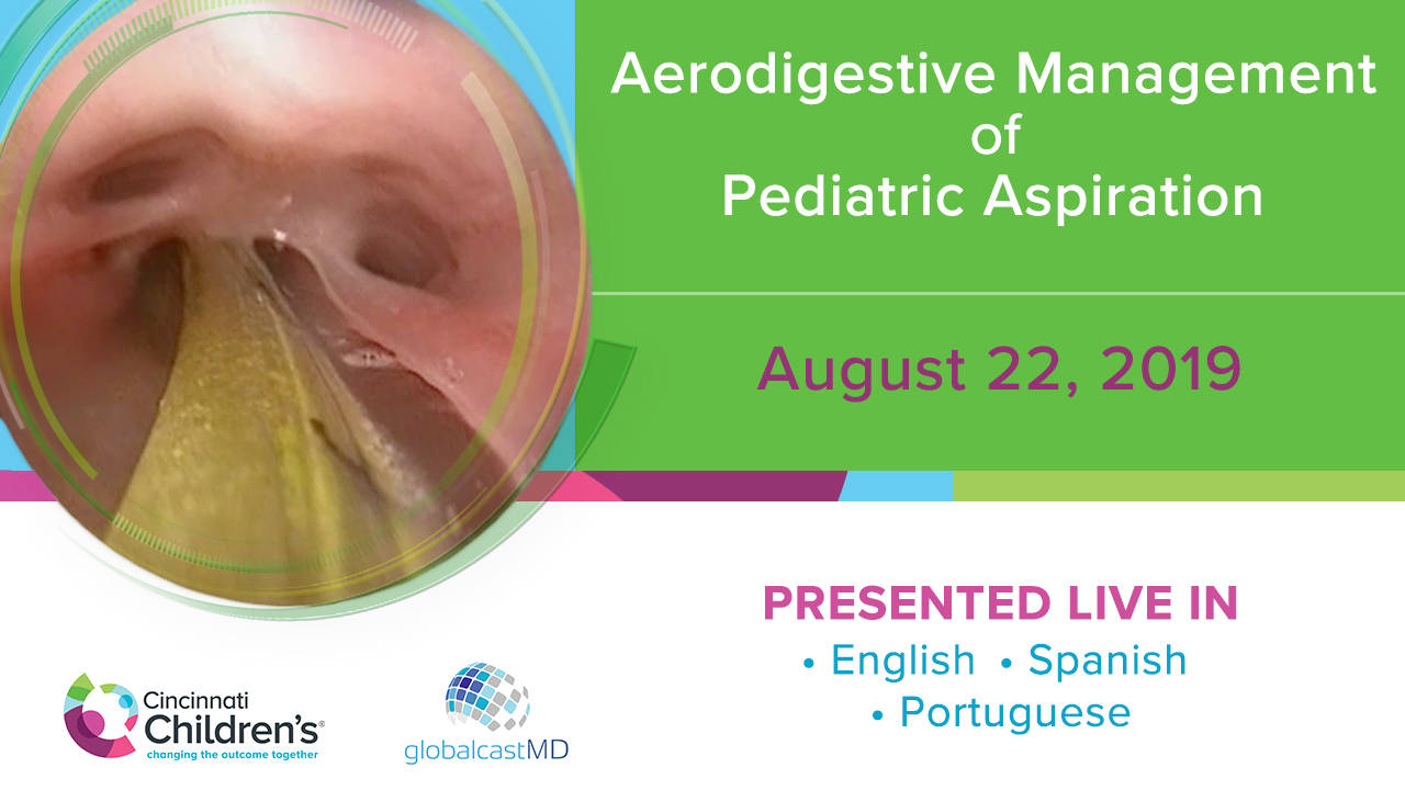
Aerodigestive Management of Pediatric Aspiration - FULL SHOW
CCHMC Pediatric Surgery · 157 min · Published Jul 2020
Video
TEF Presentations (Extended): Aerodigestive & Esophageal Surgery
108 min · Published Dec 2014
Video
QUAD #25: Medical Management of Minor Laryngeal Clefts with Dr. Greg Burg
CCHMC Pediatric Surgery · 7 min · Published Feb 2025
Video
Aerodigestive & Esophageal Surgery: Aspiration in TEFs
5 min · Published Dec 2014
Video
Surgeon annual volume impacts recurrence rates of pediatric inguinal hernia repairs: A multi-institutional study
54 s · Published Feb 2026
Podcast
Journal of Pediatric Surgery Article Review: 2nd Quarter (Apr-Jun) 2025
16 min · Published Aug 2025
Video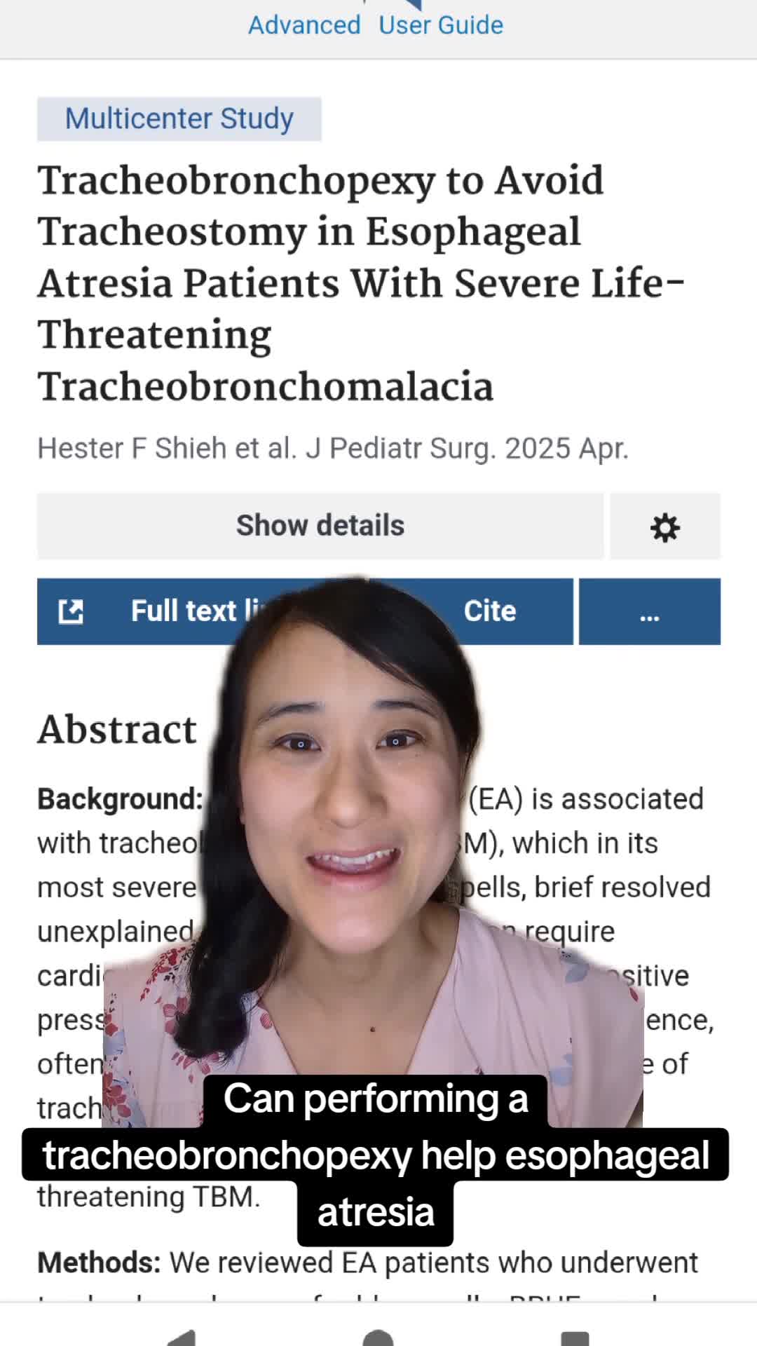
Tracheobronchopexy to Avoid Tracheostomy in Esophageal Atresia Patients With Severe Life-Threatening Tracheobronchomalacia
50 s · Published Apr 2025
Video
QUAD #27 - Multidisciplinary - How Do Teams Enhance Outcomes by the CCHMC ADEC Team
CCHMC Pediatric Surgery · 10 min · Published Apr 2025
Video
QUAD #22: What is CHARGE syndrome? with Dr. Catherine Hart
CCHMC Pediatric Surgery · 59 s · Published Dec 2024
Video
QUAD #5 Pharyngeal Scar Management with Dr. Doug von Allmen
CCHMC Pediatric Surgery · 7 min · Published Mar 2024
Video
Intestinal Atresia Types Explained: Grossfeld Classification for Pediatric Surgery
1 min · Published Sep 2026
Video
Derivation and validation of the Pediatric Community-Acquired Pneumonia Severity (PedCAPS) score: A prospective cohort study
50 s · Published Sep 2026
Video
FETO for Late-Diagnosed Severe Congenital Diaphragmatic Hernia (CDH) at Cincinnati Children's with Dr. Beth Rymeski
4 min · Published Sep 2026
Video
Severe Congenital Diaphragmatic Hernia (CDH) Case: FETO Management & Unexpected Findings with Dr. Beth Rymeski
3 min · Published Sep 2026
Video
Fetoscopic Endoluminal Tracheal Occlusion (FETO) Procedure: Step-by-Step Guide with Dr. Beth Rymeski
4 min · Published Sep 2026
Video
Choledochal Cyst Types Explained: Pediatric Surgery Fundamentals
1 min · Published Sep 2026
What the experts said
KTP laser or CO2 laser can be used for broad mucosal removal in laryngeal cleft repair, and using laser is easier to use and easier to teach with.
A laryngeal cleft is a congenital condition in which the posterior wall of the laryngotracheal tract is open and food or liquids can pass from the esophagus into the trachea, leading to aspirations.
Type 1 laryngeal cleft means the opening is above the vocal cords.
Type 2 laryngeal cleft means it extends below the vocal cords.
Type 3 laryngeal cleft means it extends down into the trachea.
The Benjamin Inglis classification should be modified to include a type 4 long because type 4 could be proximal above the carina, at the carina, or go straight through the carina.
Flexible bronchoscopy is not adequate for diagnosing a posterior laryngeal cleft.
The mass closure technique developed in Cincinnati uses the same concept as endoscopic tracheoesophageal fistula repair: you want raw against raw, with wide strips of raw tissue opposed after removing the non-stick mucosal surface.
Open approach for laryngeal clefts is reserved for failed endoscopic repair cases, some type 2s and type 3s, using a laryngofissure approach.
For type 4 clefts, a cervical approach is used.
Type 4 long clefts present anesthetic challenges requiring double lumen tube, single lung ventilation, ECMO or bypass, and often have associated microgastria and multiple congenital anomalies.
Type 4 long clefts have a very high mortality rate of approximately 50%.
The Cincinnati team does not distinguish between anatomical type 1 cleft versus deep notch for treatment decisions; they care whether the patient is aspirating (physiological cleft).
Most type 3 clefts are attempted endoscopically unless there is a reason to go open.
The transtracheal technique for type 4 clefts involves forming layers between trachea and esophagus, sewing the esophageal layer with knots in lumen, sewing the tracheal layer with knots in lumen, with optional interposition graft.
The novel surgical technique for type 4 long clefts involves transecting the trachea at the lower border of the cricoid, peeling the trachea off the esophagus beyond the cleft while keeping the patient intubated into one bronchus, repairing the esophagus with optional second imbricating layer, placing an interposition graft (typically sternal or tibial periosteum), reconnecting the trachea to the cricoid, and placing a tracheostomy relatively late at two or three weeks.
For the type 4 long cleft case, the team waited three months until the child was greater than 5 kilograms based on outcomes data from previous research.
When placing an endotracheal tube for type 4 long cleft repair, think big, like 4.5 size.
The biggest risk with laryngotracheal esophageal clefts is that the distal end turns into a tracheoesophageal fistula.
Residual tracheoesophageal fistula can be repaired endoscopically using bugbee cautery to demucosalize the tract followed by endoscopic suture placement with P2 needle on 4-0 PDS to create raw-against-raw closure.