

IPEG 2018 - RIGHT THORACOSCOPIC REPAIR OF AN H-TYPE TRACHEOESOPHAGEAL FISTULA
With Dr. Steve Rothenberg
Chapter 1 of 4 · Case-Based Learning
Case presentation
Case presentation and diagnosis
This video is for verified healthcare professionals.Sign in to watch — the rest of this page is open.Sign in
Expert statements on this page
No expert statements were drawn from this page.
Host summaries · secondary, not cited in answers
Educational content from recorded physician discussions — not medical advice. Talk to your (or your child's) care team about your situation.
Video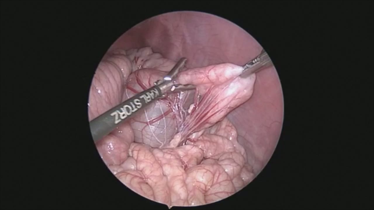
Laparoscopic Mesenteric and Short Gastric Dissection with a Vessel Sealer
Dr. Steve Rothenberg · 3 min · Published May 2026
Video
Laparoscopic Pyloromyotomy with a Reusable Handle and Disposable Blade
Dr. Steve Rothenberg · Published May 2026
Video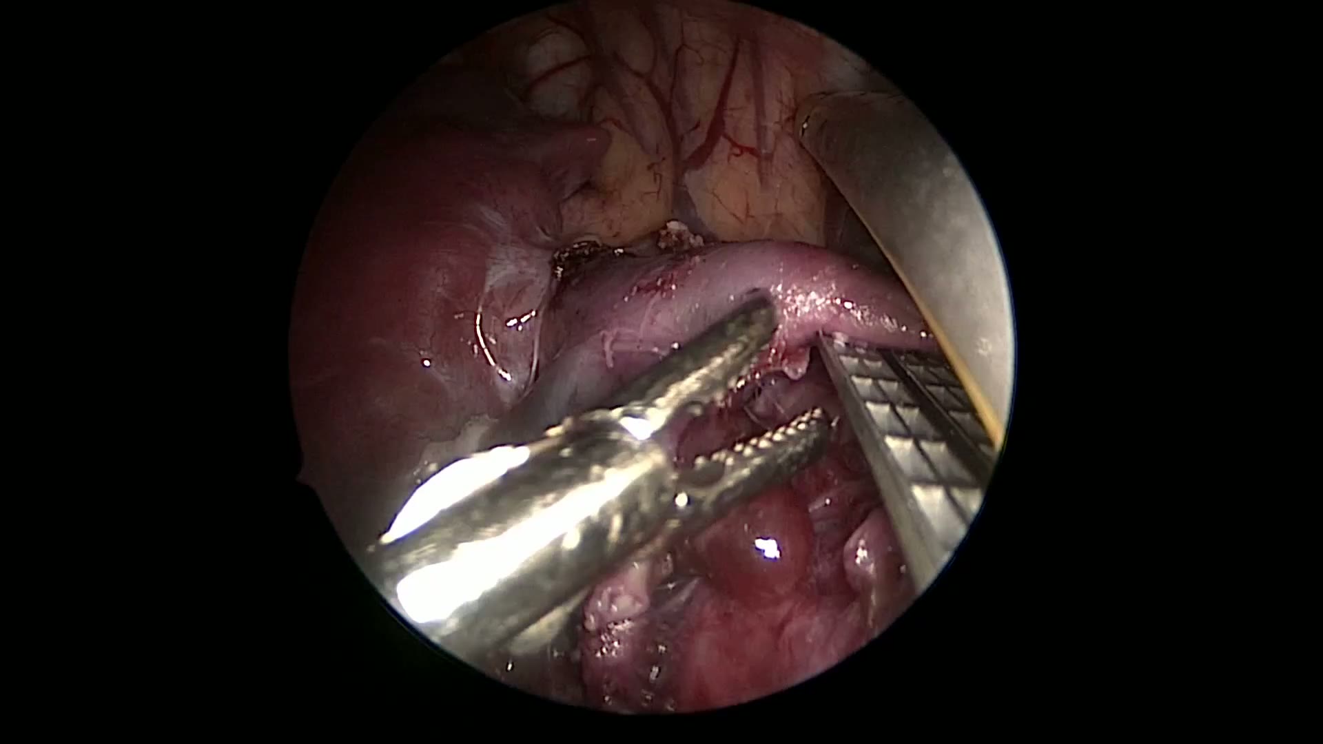
Thoracoscopic Stapling of the Left Lower Lobe Pulmonary Artery with a 5 mm Stapler
Dr. Steve Rothenberg · 9 s · Published May 2026
Video
Laparoscopic Plication of a Right Diaphragmatic Eventration in a 3-Month-Old
Dr. Steve Rothenberg · 6 min · Published May 2026
Video
Laparoscopic Redo Nissen Fundoplication in a 30 kg Child with the 3 mm Percuvance System
Dr. Steve Rothenberg · 5 min · Published May 2026
Video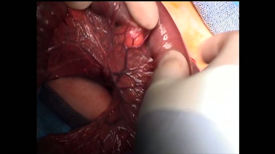
Open Bowel Anastomosis
Dr. Steve Rothenberg · Published May 2026
What the experts said
H-type TEFs are very rare and represent about 4% of all TEFs
H-type TEFs are usually diagnosed later in infancy when the patient presents with difficulty swallowing, aspiration, and recurrent pneumonias
The procedure was performed using 3 ports: a camera port behind the tip of the scapula at approximately the 4th intercostal space, a working port directly below the camera port, and another one anterior to it at the mid-axillary line
The parietal pleura at the groove of the esophagus and the trachea was opened using a combination of blunt dissection and a bipolar energy device
A 5 millimeter stapler was used to transect the tracheoesophageal fistula
A pleural flap was placed between the staple lines to help avoid a recurrent fistula
The patient had a swallow study on post-op day 2 that was negative for a leak
Advantages to a thoracoscopic H-type TEF repair are direct and magnified visualization between the esophagus and the trachea
Thoracoscopic approach allows visualization of the vagus nerve and avoidance of injury to the recurrent laryngeal nerve
Thoracoscopic approach avoids a neck incision