


Chapter 1 of 3 · Surgical Management
Setup & ports
Patient positioning and port placement
Expert statements on this page
No expert statements were drawn from this page.
Host summaries · secondary, not cited in answers
Educational content from recorded physician discussions — not medical advice. Talk to your (or your child's) care team about your situation.
Video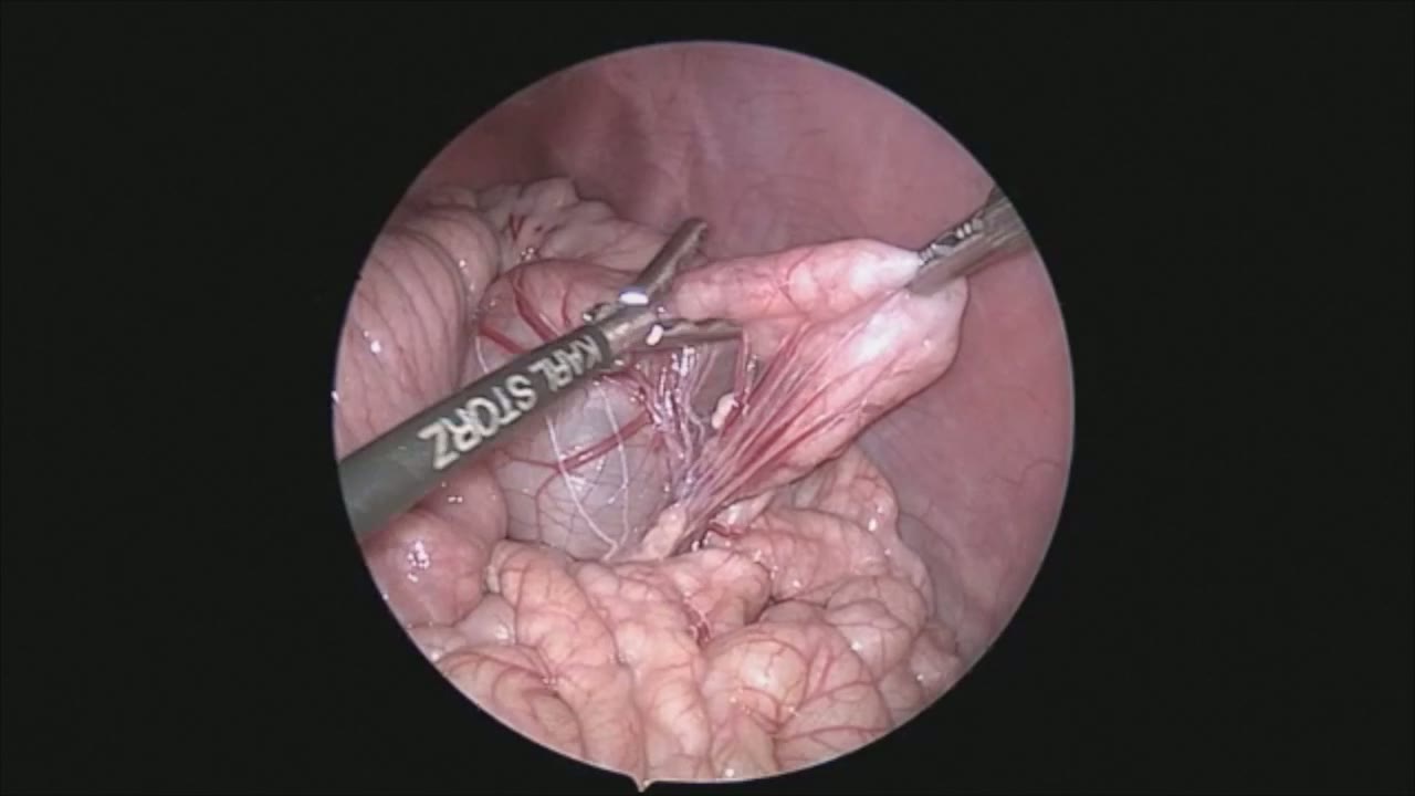
sealer demo
Dr. Steve Rothenberg · 3 min · Published May 2026
Video
pyloric knife
Dr. Steve Rothenberg · Published May 2026
Video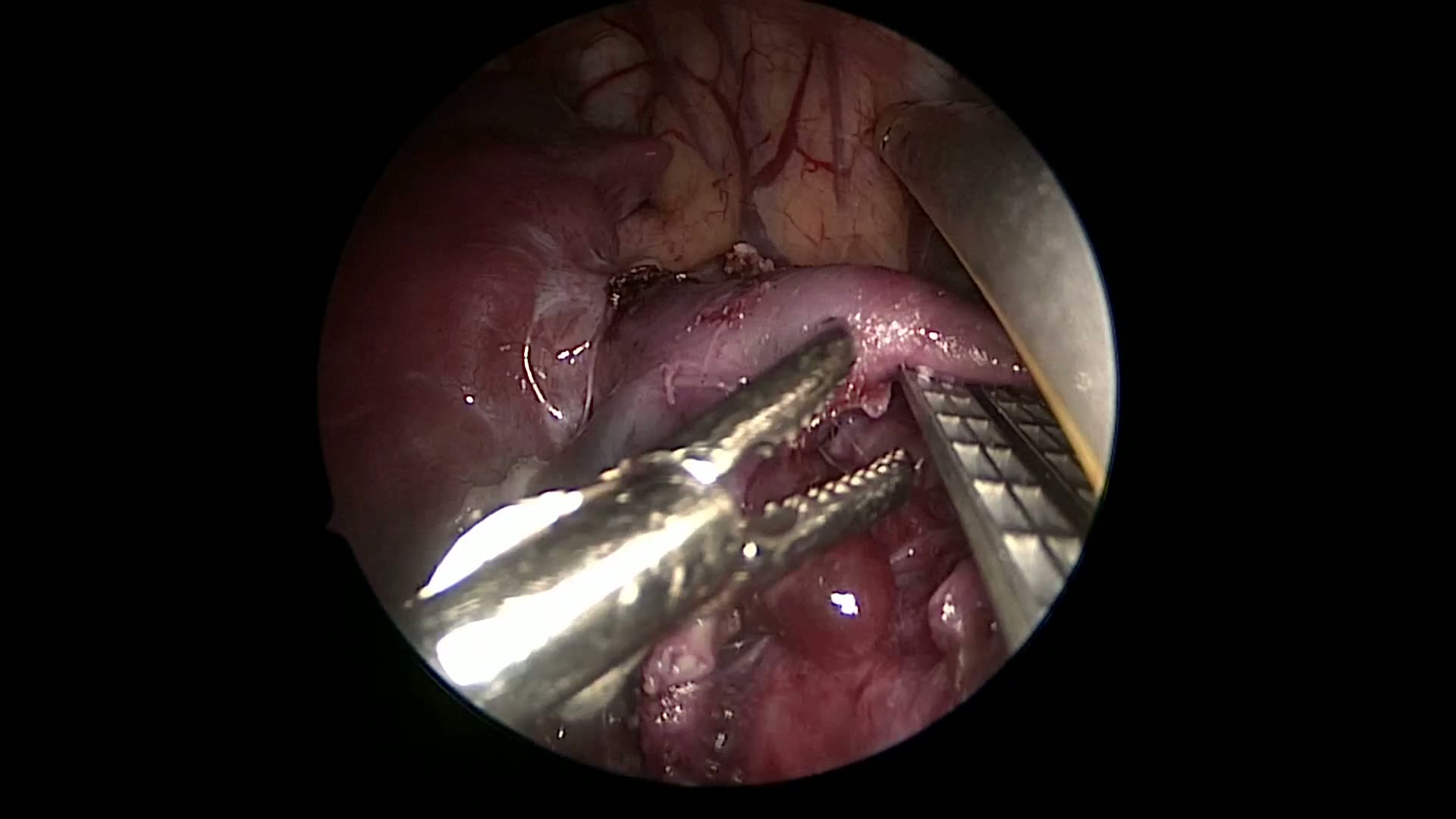
pulm artery LLL
Dr. Steve Rothenberg · 9 s · Published May 2026
Video
plication narrated
Dr. Steve Rothenberg · 6 min · Published May 2026
Video
percuvance redo nissen
Dr. Steve Rothenberg · 5 min · Published May 2026
Video
open bowel anast
Dr. Steve Rothenberg · Published May 2026
Video
Intestinal Atresia Types Explained: Grossfeld Classification for Pediatric Surgery
1 min · Published Sep 2026
Video
Derivation and validation of the Pediatric Community-Acquired Pneumonia Severity (PedCAPS) score: A prospective cohort study
50 s · Published Sep 2026
Video
FETO for Late-Diagnosed Severe Congenital Diaphragmatic Hernia (CDH) at Cincinnati Children's with Dr. Beth Rymeski
4 min · Published Sep 2026
Video
Severe Congenital Diaphragmatic Hernia (CDH) Case: FETO Management & Unexpected Findings with Dr. Beth Rymeski
3 min · Published Sep 2026
Video
Fetoscopic Endoluminal Tracheal Occlusion (FETO) Procedure: Step-by-Step Guide with Dr. Beth Rymeski
4 min · Published Sep 2026
Video
Choledochal Cyst Types Explained: Pediatric Surgery Fundamentals
1 min · Published Sep 2026
What the experts said
Single lung ventilation is generally not required for thoracoscopic diaphragmatic plication
CO2 is used to compress the lung as well as help compress the diaphragm during the procedure
A knot pusher is generally used to tie the reefing sutures down because of the tension and the small spaces and difficult angles
At least 3 bites of the diaphragm are taken with each stitch, which helps roll the diaphragm in and flatten it out
In general, 5 or 6 sutures are placed in line for the initial set of plication sutures
With each suture, more room develops within the chest cavity and makes the placement of further sutures easier
Care should be taken not to apply too much tension on any one suture as these stitches can tear through the diaphragmatic muscle
A knot pusher works well because the tissues are relatively strong and there is a fair amount of tension while trying to flatten the diaphragm
A second row of sutures is placed, imbricating the first row, to bring in more tissue and decrease the laxity in the diaphragm
In general, you will lose about 10 to 15% of your repair after the first few weeks as measured on chest X-ray, but this is not usually significant
A chest tube is generally not left after this procedure
Patients can often be weaned quickly from the ventilator and extubated after thoracoscopic diaphragmatic plication