
Wilms Tumor: Audio Chapter
With Dr. Peter Ehrlich · hosted by Dr. Em Gootee & Dr. Todd Ponsky & Dr. Abdul Raoof Lamoshi · StayCurrentMD
Part of
Wilms Tumor 20 items
Cued at 1:00:29 · stops at 1:01:14 · press play
Educational content from recorded physician discussions — not medical advice. Talk to your (or your child's) care team about your situation.
Podcast
Wilms Tumor: Audio Chapter
64 min · Published Jun 2017
Video
Wilms Tumor Protocol Violations: Practice Gap discussion at Update Course 2018
9 min · Published Aug 2018
Video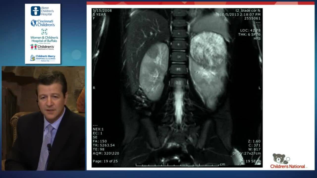
Wilms Tumor Rapid Fire: Update Course 2015
8 min · Published Nov 2015
Podcast
Topics in 10: Wilms Tumor
12 min · Published Aug 2019
Video
Wilms Tumor
Published May 2020
Podcast
Neuroblastoma
56 min · Published Aug 2019
Only a few other public items share this expert — go deeper there →
Video
Pediatric Surgical Oncology Research Collaborative (PSORC): Studying Rare Pediatric Tumors
56 s · Published May 2026
Video
Update Course Rewind 2025: Hirschsprung + ARM: Rare but Real
1 min · Published May 2026
Video
Pooling Patients to Study Rare Pediatric Tumors: An Introduction to PSORC
56 s · Published May 2026
Video
The fetal frontier: A review of current and emerging fetal therapies for genetic diseases
44 s · Published May 2026
Video
Indocyanine green assists with sentinel lymph node mapping in pediatric and adolescent patients
1 min · Published May 2026
Video
Tricuspid valve surgery in transposition of the great arteries with a systemic right ventricle
46 s · Published May 2026
What the experts said
Wilms tumors characteristically show a 'claw sign' on imaging where normal kidney is displaced into a horseshoe pattern, appearing to grab the mass coming out of it.
Wilms tumor tends to push structures out of the way rather than growing around them, whereas neuroblastoma grows around structures like blood vessels.
In North America, the Children's Oncology Group recommends primary nephrectomy and ureterectomy with lymph node sampling for the majority of children with renal tumors.
Preoperative chemotherapy is recommended if the tumor compromises the child's respiratory status, making them a poor operative candidate.
If tumor extends into the inferior vena cava beyond the intrahepatic level (behind the liver or up to the atrium), preoperative chemotherapy is recommended.
Massive tumors that would require resection of large parts of liver or bowel should receive preoperative chemotherapy because the majority will respond.
If the child has only one functioning kidney, preoperative chemotherapy is recommended to avoid nephrectomy.
Children with bilateral renal tumors, Wilms tumor predisposition syndromes (Wagr, Denys-Drash, Beckwith-Wiedemann), or multicentric tumors should not undergo primary nephrectomy.
Complication rates are higher if liver or bowel must be resected at the same time as the kidney.
If gross tumor is left behind or the tumor is only biopsied, it is treated as a stage 3 abdominal tumor requiring 3-drug chemotherapy plus flank radiation.
In the COG staging system, treatment is determined by both local abdominal stage and disease stage.
Stage 1 tumor is limited to the kidney, completely resected, with no capsular invasion, no rupture or biopsy prior to removal, no renal sinus vessel involvement, negative margins, and negative regional lymph nodes.
Stage 2 tumor is completely resected with negative margins but extends beyond the kidney through capsular penetration, renal sinus soft tissue invasion, or blood vessel involvement outside the primary kidney.
Stage 3 includes tumors that are biopsied with gross residual, have positive lymph nodes, penetrate the peritoneal surface with implants, have positive margins, have microscopic residual from intraoperative spill, cannot be completely resected, or must be removed in pieces.
Stage 4 is hematogenous metastasis to lung, liver, bone, or brain.
Stage 5 is bilateral renal tumor involvement.
Patients with stage 1 or 2 abdominal disease without lung metastases receive only 2-drug chemotherapy (vincristine and dactinomycin) for shorter duration with lower toxicity and significantly lower risk of late effects.
The main late effects of Wilms tumor treatment are renal failure, second malignancies, pregnancy problems in females, hypertension, and cardiovascular disease, primarily caused by radiation and doxorubicin.
If a child has stage 1 or 2 abdominal disease, they do not require abdominal radiation regardless of lung metastases status.
Fifteen percent of girls who receive pulmonary radiation for Wilms tumor develop breast cancer.
Pulmonary radiation causes pneumonitis and long-term restrictive lung disease.
A recent COG study showed that approximately 40% of patients with pulmonary metastases who achieve complete response by 6 weeks of chemotherapy do not need pulmonary radiation, with 80-85% remaining relapse-free.
In the SIOP protocols, all patients start with chemotherapy without biopsy in most cases, using higher doses of two drugs, with evaluation at 4 and 8 weeks before proceeding to resection.
SIOP uses post-chemotherapy, post-nephrectomy classification into low risk, intermediate risk, and high risk based on percentage of blastemal components and presence of anaplasia.
Some children with Wilms tumor develop acquired von Willebrand disease; in the majority of cases it is meaningless, but a few case series report significant bleeding during surgery until the tumor is removed.
Positive surgical margins or tumor rupture (intraoperative or microscopic) automatically makes the patient stage 3.
Taking a rim of diaphragm or liver capsule to avoid tumor violation does not upstage the tumor if the tumor itself is not divided.
Very low-risk patients (stage I, <550g, <2 years, favorable histology) can be treated with surgery alone; 90-95% are cured without chemotherapy, and those who relapse have 100% survival with delayed chemotherapy.
IVC tumor extension is not a negative prognostic factor if the tumor is completely resected.
If IVC tumor thrombus extends into the renal vein but is not adherent and comes out in one piece, it is considered stage 2.
Major complication rates (including mortality, blood transfusions, ICU stay) increase significantly when tumor extends beyond the infrahepatic IVC to the hepatic veins or atrium, with 26-30% major morbidity in primary resection cases.
Loss of heterozygosity at both 1p and 16q occurs in 5-7% of patients and is associated with significantly worse outcomes regardless of stage.
Stage 1-2 patients with loss of heterozygosity at 1p and 16q have approximately 10% lower overall survival than those without these genetic changes.
Stage 3-4 patients with loss of heterozygosity at 1p and 16q have approximately 18% lower overall survival and receive 5-drug regimen M chemotherapy.
Bilateral Wilms tumors occur in 8-10% of all children with Wilms tumor.
Event-free survival for unilateral Wilms tumor is approximately 88% with overall survival of 95%.
Event-free survival for bilateral Wilms tumor on NWTS-5 was 61% with overall survival of only 80%.
The maximum response of most children with Wilms tumor to chemotherapy occurs by 12 weeks.
In children presenting under 36 months with bilateral renal tumors, it is almost universally Wilms tumor.
Open biopsy to determine favorable vs. unfavorable histology in Wilms tumor is not very accurate initially.
In a recent COG bilateral Wilms study of 250 patients, only one patient who met enrollment criteria turned out to have rhabdoid tumor instead of Wilms tumor.
Discordant pathology occurs in up to 20% of bilateral Wilms tumor patients when both kidneys are biopsied.