
Biliary Atresia Part I
With Dr. Greg Tiao & Dr. Atsuyuki Yamataka & Dr. Mark Davenport · hosted by Dr. Todd Ponsky · StayCurrentMD
Part of
Biliary Atresia 26 items
Cued at 15:47 · stops at 16:32 · press play
Educational content from recorded physician discussions — not medical advice. Talk to your (or your child's) care team about your situation.
Video
Biliary Atresia
CCHMC Pediatric Surgery · 20 min · Published Nov 2018
Video
Biliary Atresia - Clinical Practice Updates
Published Dec 2020
Video
Biliary Atresia: Update Course 2015
CCHMC Pediatric Surgery · 20 min · Published Jul 2017
Video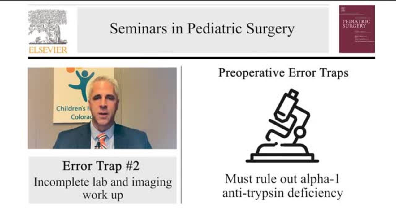
Error Traps and Culture of Safety in Biliary Atresia
CCHMC Pediatric Surgery · 4 min · Published Nov 2019
Podcast
Biliary Atresia Part II
42 min · Published Feb 2022
Video
Biliary Atresia-Case Presentation and Panel Discussion: Update Course 2014
23 min · Published Jul 2017
Video
Update Course Rewind: MMP-7 & Biliary Atresia Diagnosis 2024
CCHMC Pediatric Surgery · 6 min · Published Mar 2025
Video
Biliary Atresia with Dr. Greg Tiao
CCHMC Pediatric Surgery · 11 min · Published Jun 2022
Video
Hepatoblastoma with Dr. Greg Tiao
CCHMC Pediatric Surgery · Published Mar 2022
Podcast
Hepatoblastoma with Dr. Greg Tiao
10 min · Published Feb 2022
Video
Compiled Sandler Rapid Fire Sessions: Update Course 2015
29 min · Published Nov 2015
Video
Pediatric Surgical Oncology Research Collaborative (PSORC): Studying Rare Pediatric Tumors
56 s · Published May 2026
Video
Update Course Rewind 2025: Hirschsprung + ARM: Rare but Real
1 min · Published May 2026
Video
Update Course Rewind 2025: Hirschsprung + ARM: Rare but Real
1 min · Published May 2026
Video
Pooling Patients to Study Rare Pediatric Tumors: An Introduction to PSORC
56 s · Published May 2026
Video
The fetal frontier: A review of current and emerging fetal therapies for genetic diseases
44 s · Published May 2026
Video
Indocyanine green assists with sentinel lymph node mapping in pediatric and adolescent patients
1 min · Published May 2026
What the experts said
A high GGT is more consistent with an obstructive process and helps guide toward eliminating PFIC (progressive familial intrahepatic cholestasis) diagnoses.
At 50 days of age, physiologic jaundice from the newborn period would have resolved.
The key diagnostic finding is conjugated hyperbilirubinemia.
Infants who initially had yellow stool that became clay-colored have better prognosis than those with clay-colored stool immediately after passing meconium.
Ultrasound separates out other surgical issues including inspissated bile syndrome and spontaneous perforation of bile ducts, which should have ultrasound-positive features.
The triangular cord sign on ultrasound is not particularly discriminatory for biliary atresia.
Cincinnati Children's Hospital has not used HIDA scans for biliary atresia diagnosis in approximately 25 years.
Experienced pathologists can diagnose biliary atresia from percutaneous liver biopsy in almost 90% of cases.
King's College Hospital uses ERCP for approximately 10% of biliary atresia cases when biopsy findings are equivocal.
Histologic findings diagnostic of biliary atresia include ductular proliferation, small cell infiltrate, and bile duct plugs in proliferating bile ducts.
By 50 days of age, all histologic features of biliary atresia should be established on biopsy, whereas earlier biopsies (10-20 days) may not show consistent diagnostic findings.
Significant fibrosis on liver biopsy reflects disease progression and may guide surgical decision-making.
Liver exteriorization during Kasai portoenterostomy can create denser, more vascularized adhesions that complicate future liver transplantation.
Liver exteriorization with a small incision can cause kinking of hepatic veins.
Recent data from Juntendo shows liver function might be worse in the midterm follow-up for patients who had laparoscopic Kasai compared to open surgery.
The only chance a child with biliary atresia has to avoid early transplant is a well-done Kasai portoenterostomy.
Even in the best hands, long-term drainage rate after Kasai portoenterostomy is only 70-75%.
Damage to hepatic artery branches during Kasai dissection can exacerbate the underlying liver disease process.
The hilar plate should be transected at the level of Glisson's capsule, leaving it intact, without cutting into the liver parenchyma itself.
In the Children's Network surgical committee, approximately one-third of centers cut into the liver during Kasai, while the rest transect at 1-2 mm of fibrous remnant.
Coring into the liver creates an unstable interface that has never achieved good results in the literature, as biliary ductules that transgress simply scar over.
Standard Roux limb length for Kasai portoenterostomy is 40 centimeters, though Cincinnati uses 35-40 cm and measures with silk suture to 45 cm.
The Roux anastomosis suture choice does not make a difference in outcomes.
For the portoenterostomy anastomosis, 6-0 Maxon suture is used with all knots tied on the outside.
Yamataka uses a shorter Roux limb than standard because he does not like redundant Roux loops.
Biliary atresia is the leading cause for liver transplant in the United States.
NASPGHAN guidelines recommend not obtaining HIDA scans because phenobarbital loading delays treatment by 5-6 days, pushing closer to the window where Kasai efficacy deteriorates.
IPEG placed a moratorium on laparoscopic Kasai portoenterostomy because of poor outcomes.
Peterson ran a prospective randomized trial comparing laparoscopic to open Kasai and stopped the trial early.
Dr. Neo published a paper describing different depths of hilar plate transection in Kasai portoenterostomy.